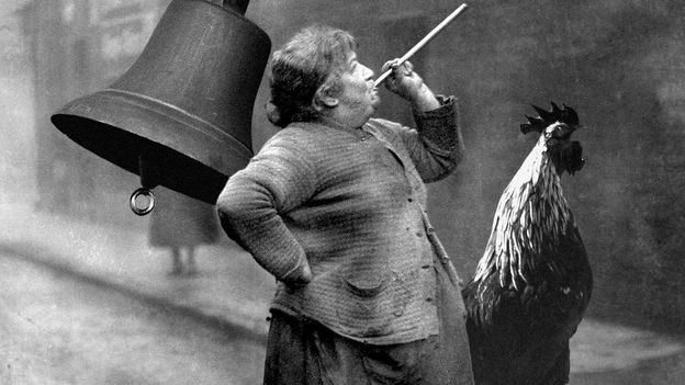


In a groundbreaking scientific endeavor years in the making, a team of researchers in Melbourne has been granted unprecedented access to study the 13 youngest preserved Tasmanian tigers in existence. These rare specimens, sourced from museums across Australia and Charles University in Prague, have been preserved for over a century and were recently rediscovered. The study represents a significant milestone, as these preserved creatures rarely leave their respective institutions.
Transporting the specimens was a delicate operation, underscoring their value. The Tasmanian Museum and Art Gallery, for instance, assigned a curator to personally escort them across the Bass Strait. The key to convincing these museums to part with their treasures was the innovative method of analysis proposed by the researchers: computed tomography (CT) scanning. This non-invasive technique, a staple in the medical field since the late 1970s, allows scientists to examine the internal structures of specimens without destroying them.
Revolutionizing Specimen Analysis with CT Scanning
CT scanning has revolutionized the way researchers can study preserved specimens. Unlike traditional methods that require physical dissection, CT scanning provides a detailed view inside the creature’s skin, skeleton, and organs. This technology has been used to scan everything from tiny meteorites to predict climate impacts on animal habitats.
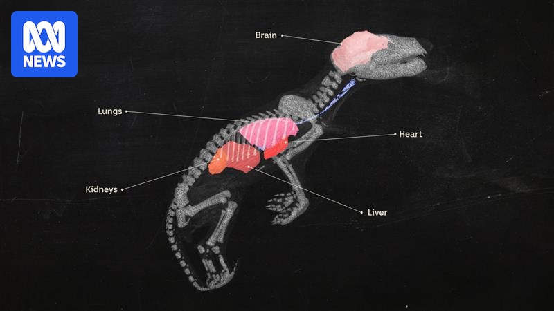
In the case of the Tasmanian tiger, the CT scans revealed intricate details of the young thylacines at various stages of development. Axel Newton, a researcher involved in the project, utilized a process known as segmentation to identify vital organs such as the heart, lungs, and brain. The scans even highlighted unexpected findings, such as the presence of two specimens that were not Tasmanian tigers but likely quolls or Tasmanian devils.
“It was absolutely magical to be able to really peel back and start to have a look at all of that incredible data that you can see when you can pick out every single tiny little facet of bone,” said Andrew Pask, an epigeneticist from the University of Melbourne.
The Broader Impact of CT Scanning in Research
The use of CT scanning extends beyond the study of Tasmanian tigers. Museums are increasingly adopting this technology because it offers high-resolution, three-dimensional copies of their specimens. These digital models can be accessed by researchers worldwide and used for educational purposes. The models can even be 3D-printed for public display, allowing people to connect with extinct species in a tangible way.
Professor Pask, who is also involved in a project to bring the thylacine back from extinction, has used CT scanning to extract DNA from preserved specimens. The success of these scans has encouraged museums to offer more specimens for analysis, highlighting the growing acceptance of this technology in the preservation and study of historical collections.
CT Scanning: A Global Trend in Museums
Globally, museums are embracing CT scanning as a means to democratize access to their collections. Dr. Christy Hipsley, a member of the Tasmanian tiger research team, has advocated for a standardized repository for CT scanned data, similar to GenBank for DNA sequences. This initiative aims to make the vast array of specimens in museum collections accessible to researchers worldwide.
In 2024, a consortium of natural history museums in the United States embarked on a project to scan and publish 13,000 vertebrate specimens, including a humpback whale. This collaborative effort underscores the potential of CT scanning to transform how museums share their collections with the world.
Future Prospects and Implications
The implications of CT scanning in research are profound. By providing a non-destructive means of studying specimens, it preserves the integrity of rare and valuable collections while offering new insights into the natural world. The ability to create detailed, digital models opens up new possibilities for research, education, and conservation efforts.
As technology continues to advance, the role of CT scanning in museums and research institutions is likely to expand. The ability to share and study digital models globally could lead to new discoveries and a deeper understanding of biodiversity and evolution. This technological shift represents a new era in the preservation and study of the world’s most precious specimens.
As museums and researchers continue to embrace CT scanning, the potential for new discoveries and a greater understanding of our natural history is immense. This innovative approach not only preserves the past but also paves the way for future scientific breakthroughs.