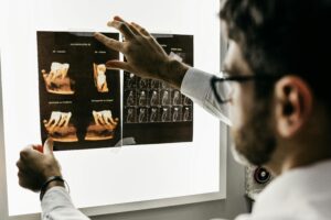
A groundbreaking study has established a link between radiation exposure from medical imaging and a small but significant risk of blood cancers in children and adolescents. Conducted by researchers at the University of Florida and funded by the National Cancer Institute, the study underscores the importance of weighing the benefits of medical imaging against its minimal risks.
Published in the New England Journal of Medicine, the paper titled “Medical Imaging and Pediatric and Adolescent Hematologic Cancer Risk” concludes that while ionizing radiation is a known carcinogen, the advantages of CT imaging in children, when justified, outweigh the potential risks. The study is expected to guide medical professionals in making informed decisions about imaging techniques for young patients.
Innovative Research Methods and Findings
Dr. Wesley Bolch, a distinguished professor in biomedical and radiological engineering at the University of Florida, played a pivotal role in the study. Using virtual patient anatomic models, Bolch and his team reconstructed bone marrow doses in over 3.7 million children who underwent CT scans between 1996 and 2016.
“We used a library of 3D anatomic whole-body computerized patient models developed in the early 2010s under a contract with the National Cancer Institute,” said Bolch, highlighting the unique methodology of the study. This approach allowed for precise measurement of leukemia risks, a significant advancement over previous models based on atomic bomb survivor data.
“This is the very first study of its kind in the U.S. and Canada, and the very first study of cancer risks in children undergoing medical imaging where each patient was considered in a unique manner regarding their sex, body size, and medical imaging exposure technique factors,” Bolch explained.
Implications for Medical Imaging Practices
The study’s findings have significant implications for the medical community. While CT imaging contributes a greater fraction of total radiation, doses from other imaging techniques like nuclear medicine, radiography, and fluoroscopy were also considered in the bone marrow-dose calculations. The highest CT doses to bone marrow were recorded in head-and-neck imaging, with an average dose of 30.8 milligray.
Despite the risks, the incidence of hematologic cancers by age 21 was only 0.3% among children exposed to bone marrow doses exceeding 30 milligray. Moreover, fewer than 1% of the 3.7 million children studied had cumulative doses above this threshold. Importantly, CT imaging doses today are significantly lower, thanks to advancements in imaging technology and faster systems.
Historical Context and Technological Advancements
This research comes 25 years after a Columbia University study linked leukemia to certain radiology scans, causing widespread concern. At that time, imaging technologists and radiologists often failed to adjust X-ray techniques for patient size, leading to unnecessary radiation exposure.
“Consider that you’ve just imaged an obese male with a high-intensity and high-energy X-ray beam, and now a petite 7-year-old girl becomes the next patient to be imaged,” Bolch explained. “In the late 1990s, very few clinics would adjust the X-ray energies and intensities from the previous adult patient. This paper scared a lot of people, but it really was a great service because it says, ‘Oh, we’re doing this wrong.'”
Since the early 2000s, there has been a concerted effort to adjust X-ray energy and intensity based on patient size, and CT system manufacturers have made significant technological improvements to reduce patient doses.
Collaboration and Future Directions
The study was led by radiologist and epidemiologist Dr. Rebecca Smith-Bindman from the University of California, San Francisco, and biostatistician Dr. Diana Miglioretti from UC Davis. Their work involved collecting and organizing medical records to determine the imaging exams performed on children and linking these to cancer registries to identify subsequent bone marrow cancers.
“This is where my laboratory came in,” Bolch said. “We ran computer simulations of these imaging procedures to provide estimates of bone marrow radiation doses for each child, for each form of medical imaging, and for each imaging examination.”
As medical imaging continues to evolve, the study emphasizes the need for ongoing research to refine imaging techniques and minimize risks. “These risks are low, and when justified by the imaging physician, patient benefits, such as disease detection, will greatly outweigh these very small risks,” Bolch concluded.
The study not only highlights the University of Florida’s leadership in making medical imaging safer for children but also sets a precedent for future research aimed at safeguarding patient health while advancing global healthcare practices.


