


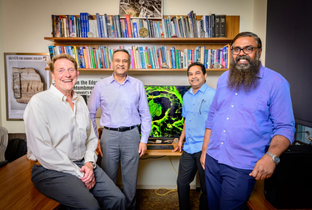
Researcher professors Bruce Fouke, Rohit Bhargava, Mayandi Sivaguru and Ashok Samuel collaborated on a paper titled, Mechanisms of osteopontin-stabilized amorphous calciumphosphate calcification in benignand pre-malignant breast disease” that hopes to guide development of methods to inhibit ACP calcifications within low-risk lesions to minimize unnecessary biopsies. Photo taken at the University of Illinois Urbana-Champaign on Tuesday, July 1, 2025. (Photo by Fred Zwicky / University of Illinois Urbana-Champaign)
Researchers at the University of Illinois Urbana-Champaign, in collaboration with the Mayo Clinic and the University of Texas at Austin, have uncovered significant structural differences between benign and cancerous calcium phosphate deposits in breast tissue. Published findings from their study suggest that these differences could lead to improved diagnostic criteria, potentially reducing the number of unnecessary biopsies and guiding future therapeutic developments.
“Dense calcifications are very common in breast tissue. They are seen easily on a mammogram, which doctors can use to classify benign, probably benign and suspicious categories,” said Bruce Fouke, study leader and professor at the University of Illinois. “But most biopsies of spots deemed suspicious end up being benign, meaning those patients underwent painful procedures unnecessarily. We want mammograms to be more precise and more accurate for distinguishing between benign breast disease and cancer.”
Breakthrough in Understanding Calcification Structures
The research team employed a multidisciplinary approach, utilizing a combination of geology, biology, and medicine, termed “GeoBioMed.” This innovative approach has previously been applied to the study of kidney stones and heart calcifications. For this study, the team examined biopsied tissue samples from benign breast disease (BBD) and ductal carcinoma in situ (DCIS), which were part of a long-term study at the Mayo Clinic.
Using a comprehensive suite of 12 different analytical techniques, including light, laser, and electron microscopy, as well as X-ray and Raman spectroscopy, the researchers documented the mineral characteristics of the samples. This detailed analysis revealed that the calcifications were composed of amorphous calcium phosphate (ACP), a mineral capable of changing shape and structure, rather than the crystalline hydroxyapatite typically found in bone.
Implications for Diagnosis and Treatment
The study found distinct differences in the formation of calcifications between BBD and DCIS. Benign calcifications tended to form spherical nodules with concentric layers, while cancerous calcifications were more elongated and irregular. Some cancerous nodules exhibited a fossilization process similar to petrified wood.
“The types of ACP nodules we saw were completely unknown and establish a brand-new classification scheme between BBD and DCIS,” Fouke explained. “Each has a different genesis and history of formation, reflecting changes in breast physiology that in turn correlated strongly with whether a biopsy sample was designated benign, possibly benign or suspicious.”
Recognizing that calcifications are composed of ACP rather than hydroxyapatite opens new treatment possibilities. Certain drugs can dissolve ACP deposits, potentially reducing misidentifications in mammograms and preventing millions of unnecessary biopsies, according to Mayandi Sivaguru, the study’s first author.
Future Directions and Collaborative Efforts
The collaboration between the University of Illinois and the Mayo Clinic exemplifies the potential for innovative research to impact public health. Rohit Bhargava, co-author of the study and director of the Cancer Center at Illinois, emphasized the unique partnerships that have facilitated this research, which could lead to improved breast cancer care.
Looking ahead, the researchers plan to extend their study to more advanced invasive breast cancer cases and explore how calcification might influence the progression of DCIS to invasive cancer. They also intend to utilize a GeoBioCell, an experimental microfluidic device developed by the Illinois team, to further study ACP calcifications.
“It’s a roadmap for the future of controlled experimental testing,” Fouke stated. “For instance, if we want to know, if a woman was to drink more water, would it make a difference in the amount or type of breast calcifications? We could flow more or less water and breast fluids through samples in the GeoBioCell and track how the calcifications grow. Similarly, we could test the effectiveness of drugs and combinations of different plant extracts to find new therapeutic agents.”
The study was supported by the Barbara and Ed Weil Foundation, with Fouke holding the Ralph E. Grim professorship at Illinois and being a member of the Cancer Center at Illinois. The findings mark a significant step forward in the understanding and potential treatment of breast calcifications, promising a future where diagnostic precision and therapeutic options are greatly enhanced.