


For individuals at high risk of developing breast cancer, frequent screenings are crucial for early detection. Researchers at the Massachusetts Institute of Technology (MIT) have unveiled a groundbreaking miniaturized ultrasound system designed to facilitate more frequent breast ultrasounds, potentially even from the comfort of one’s home or at a doctor’s office. This innovation promises to enhance early detection rates, thereby improving treatment outcomes.
The newly developed system comprises a compact ultrasound probe connected to an acquisition and processing module, slightly larger than a smartphone. This portable system, when linked to a laptop, can reconstruct and display wide-angle 3D images in real-time. According to Canan Dagdeviren, associate professor of media arts and sciences at MIT and senior author of the study, “Everything is more compact, and that can make it easier to be used in rural areas or for people who may have barriers to this kind of technology.”
The research, published in the journal Advanced Healthcare Materials, was led by Colin Marcus, PhD ’25, and former MIT postdoc Md Osman Goni Nayeem. Other contributors include MIT graduate students Aastha Shah, Jason Hou, and Shrihari Viswanath, among others.
Addressing the Need for Frequent Monitoring
While routine mammograms are a common method for detecting breast tumors, they are not foolproof. Tumors can develop between annual mammograms, known as interval cancers, which account for 20 to 30 percent of all breast cancer cases and tend to be more aggressive. Early detection is vital, as breast cancer diagnosed in its earliest stages has a survival rate nearing 100 percent, compared to around 25 percent for later-stage detections.
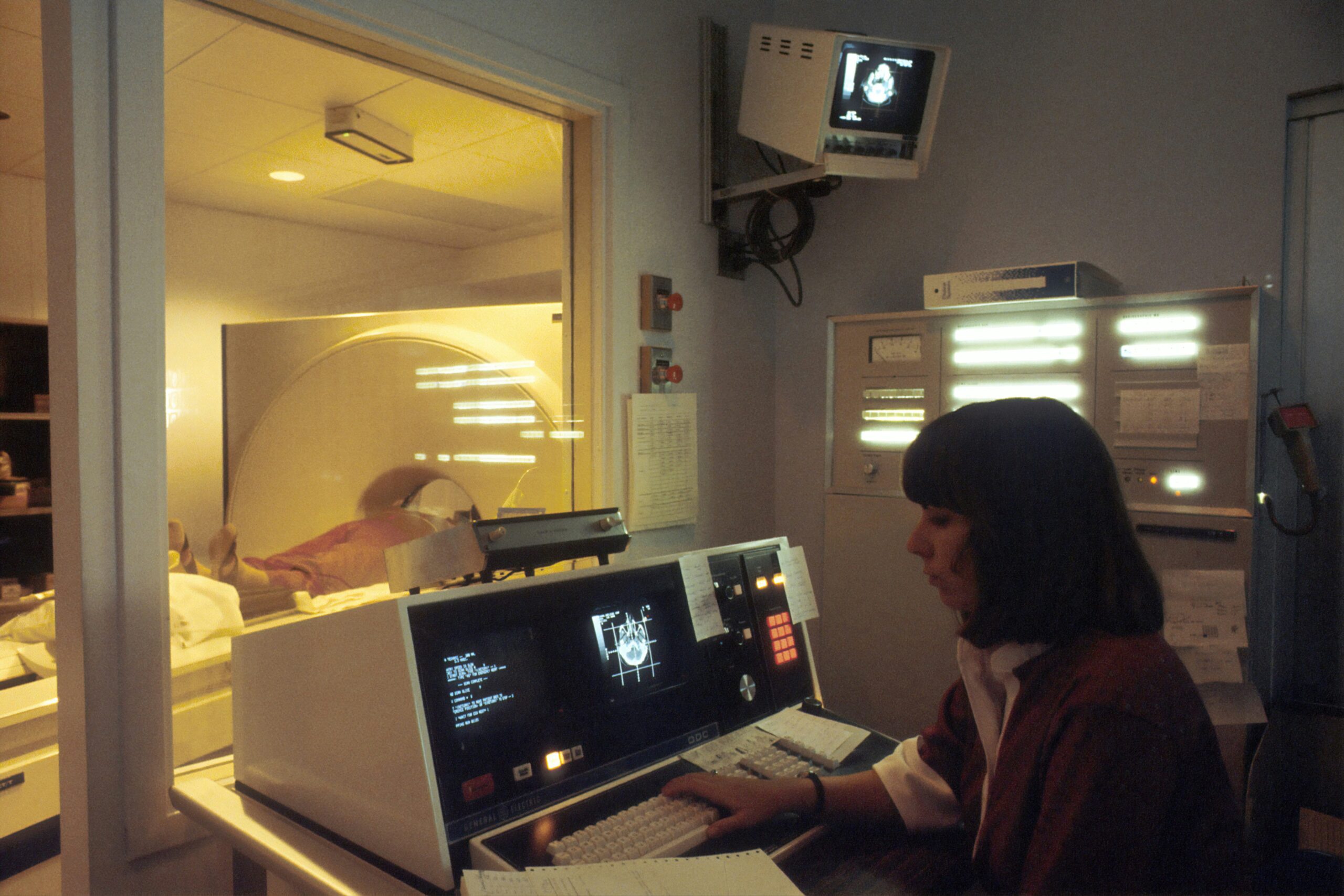
Currently, ultrasound is typically used as a follow-up to mammograms, requiring large, expensive machines and skilled technicians. “You need skilled ultrasound technicians to use those machines, which is a major obstacle to getting ultrasound access to rural communities, or to developing countries where there aren’t as many skilled radiologists,” explains Shrihari Viswanath.
The MIT team’s portable ultrasound system aims to overcome these barriers, making frequent scanning more accessible to a broader population.
Technological Innovations and Advancements
In 2023, Dagdeviren and her team developed an array of ultrasound transducers incorporated into a flexible patch, enabling users to image breast tissue from various angles. However, this system required connection to a traditional, costly processing machine, limiting its practicality.
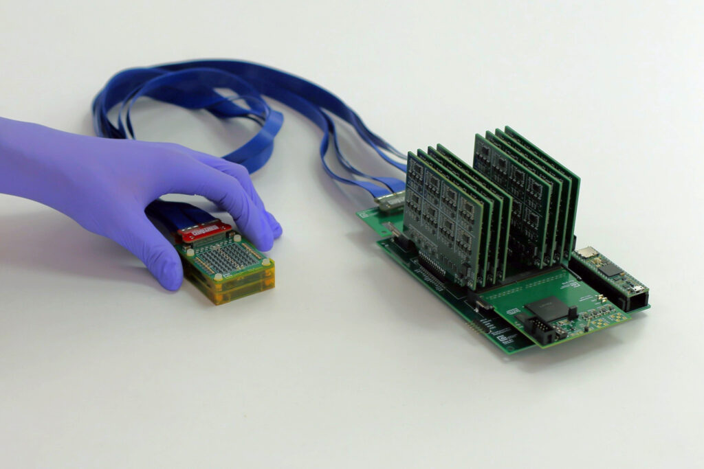
The latest study introduces a chirped data acquisition system (cDAQ), featuring an ultrasound probe and a motherboard for data processing. The probe, smaller than a deck of cards, contains an ultrasound array arranged in an empty square configuration, allowing for 3D imaging of the tissue. The motherboard, slightly larger than a smartphone, is affordable at approximately $300 and uses commercially available electronics. This setup can be connected to a laptop, making the entire system portable.
“Traditional 3D ultrasound systems require power expensive and bulky electronics, which limits their use to high-end hospitals and clinics,” says Anantha Chandrakasan, MIT Provost. “By redesigning the system to be ultra-sparse and energy-efficient, this powerful diagnostic tool can be moved out of the imaging suite and into a wearable form factor that is accessible for patients everywhere.”
The system’s low power consumption allows it to operate with a 5V DC supply, such as a battery or an AC/DC adapter, further enhancing its portability.
Clinical Trials and Future Prospects
Initial tests of the new system on a 71-year-old woman with a history of breast cysts demonstrated its efficacy in accurately imaging cysts and creating a 3D image of the tissue without gaps. The device can image up to 15 centimeters deep and cover the entire breast from just two or three locations. Unlike traditional probes, this device sits on top of the skin, preventing image distortion.
“With our technology, you simply place it gently on top of the tissue and it can visualize the cysts in their original location and with their original sizes,” Dagdeviren notes.
The research team is conducting a larger clinical trial at the MIT Center for Clinical and Translational Research and Massachusetts General Hospital. They are also working on a smaller version of the data processing system, aiming to integrate it with a smartphone for easier use. A future smartphone app could guide users on optimal probe placement using AI algorithms.
Dagdeviren is in the process of launching a company to commercialize this technology, supported by various MIT initiatives and grants. The research received funding from a National Science Foundation CAREER Award, a 3M Non-Tenured Faculty Award, the Lyda Hill Foundation, and the MIT Media Lab Consortium.
As the technology advances, the hope is to incorporate it into wearable sensors for home use, particularly benefiting those at high risk for breast cancer. This innovation represents a significant step forward in making breast cancer screening more accessible and frequent, potentially saving countless lives through earlier detection and treatment.