


In a groundbreaking study by researchers at the Massachusetts Institute of Technology (MIT), new insights have emerged into how the blurry vision of newborns may play a crucial role in shaping brain pathways. The study, published in the journal Communications Biology, explores how early visual experiences influence the development of two primary pathways in the brain’s visual system.
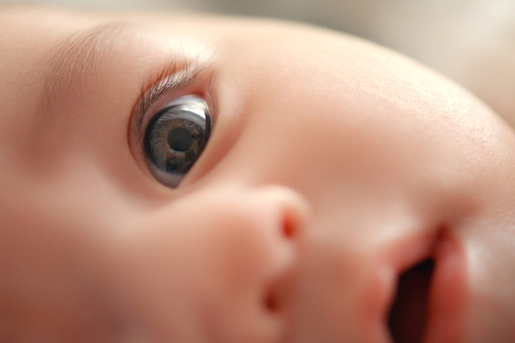
Newborns typically exhibit poor visual acuity and color vision due to underdeveloped retinal cone cells. This results in a world perceived as blurred and color-reduced. The MIT research team, led by Professor Pawan Sinha, suggests that this early visual limitation may encourage certain brain cells to specialize in processing low spatial frequencies and limited color, aligning with the magnocellular system. As vision sharpens, other cells may adapt to finer details and richer color, consistent with the parvocellular system.
Unveiling the Visual Pathways
The visual system processes information through two main pathways: the magnocellular pathway, which handles spatial localization and high temporal frequencies, and the parvocellular pathway, responsible for color and fine spatial detail. The MIT study proposes that the developmental trajectory of these pathways is influenced by the quality of visual input during infancy.
To test their hypothesis, researchers trained computational models to simulate the visual experiences of human infants. Initially, these models were exposed to low-quality images, gradually transitioning to full-color, sharper images. The outcomes revealed that models trained on this trajectory developed processing units reminiscent of the magnocellular and parvocellular pathways. In contrast, models exposed only to high-quality images lacked such distinct characteristics.
“The findings potentially suggest a mechanistic account of the emergence of the parvo/magno distinction, which is one of the key organizing principles of the visual pathway in the mammalian brain,” said Pawan Sinha, senior author of the study.
Insights from Project Prakash
The idea that low-quality visual input could benefit development is not new. It stems from studies of children who were born blind and later had their sight restored. Project Prakash, an initiative from Sinha’s laboratory, has screened and treated thousands of children in India with reversible vision loss. Post-treatment, many children participate in studies tracking their visual development.
In one such study, children who had cataracts removed showed a significant drop in object-recognition performance when presented with black and white images compared to colored ones. This led researchers to hypothesize that reduced color input during early development might help the brain learn to recognize objects even in images with impoverished colors.
“Denying access to rich color at the outset seems to be a powerful strategy to build in resilience to color changes and make the system more robust against color loss in images,” Sinha explained.
Modeling Developmental Progressions
Building on these findings, the MIT team explored the consequences of limited color and visual acuity at the start of development. They hypothesized that these limitations could contribute to the development of the magnocellular and parvocellular pathways. To test this, they trained models on two different sets of images: one standard dataset and another biomimetic dataset mimicking human visual input from birth.
The biomimetic data consisted of low-resolution, grayscale images followed by high-resolution, colorful images. Analysis revealed that models trained on this data developed distinct units responsive to low-color and low-spatial-frequency inputs, akin to the magnocellular pathway. Additionally, they exhibited parvocellular-like units tuned to higher spatial frequencies or richer color signals, a distinction absent in models trained on high-quality images from the start.
“This provides some support for the idea that the ‘correlation’ we see in the biological system could be a consequence of the types of inputs that are available at the same time in normal development,” said Lukas Vogelsang, a lead author of the study.
Implications for Understanding Brain Organization
Further experiments revealed that models trained on biomimetic input were more likely to use an object’s shape for categorization, similar to human perception. When magnocellular-like units were removed, the models lost this tendency. Moreover, models trained on videos showed that units tuned to high temporal frequencies also exhibited magnocellular-like properties, suggesting a link between early visual input and the organization of sensory processing pathways.
Overall, the study supports the notion that low-quality sensory input early in life may contribute to the organization of brain pathways. While it does not rule out innate specifications, it provides evidence that visual experience during development plays a significant role.
“The general theme that seems to be emerging is that the developmental progression we go through is very carefully structured to give us certain kinds of perceptual proficiencies, and it may also have consequences in terms of the very organization of the brain,” Sinha concluded.
This research was funded by the National Institutes of Health, the Simons Center for the Social Brain, the Japan Society for the Promotion of Science, and the Yamada Science Foundation.