


Seventy years ago, Erwin W. Müller, a pioneering physicist at Penn State, made history by becoming the first person to “see” an atom. This groundbreaking achievement took place in the Osmond Laboratory on Penn State’s University Park campus, solidifying Müller’s legacy as a trailblazer in the field of physics and beyond. His work laid the foundation for advancements in atomic resolution imaging, a cornerstone of modern microscopy.
Müller, originally from Germany, joined Penn State’s faculty in 1951 when the institution was known as the Pennsylvania State College. His research was conducted in the field emission laboratory, initially located in the sub-basement of Osmond, before moving to the second floor in 1954. Over two decades, Müller focused on enhancing the resolution of microscope images, leading to significant technological breakthroughs.
The Journey to Atomic Resolution
In 1936, Müller invented the field emission microscope, a device that nearly achieved atomic resolution by studying the surfaces of needle tips. He continued his work by developing the field ion microscope in 1951, which ultimately allowed him to “see” tungsten atoms in 1955. This was a pivotal moment in microscopy.
Kanwar Bahadur, Müller’s graduate student, played a crucial role in this discovery. In 1955, Bahadur experimented with cooling the tungsten tip of the field ion microscope using liquid nitrogen, aiming to enhance resolution. His adjustments led to a breakthrough, prompting Müller to exclaim, “Atoms, ja, atoms!”
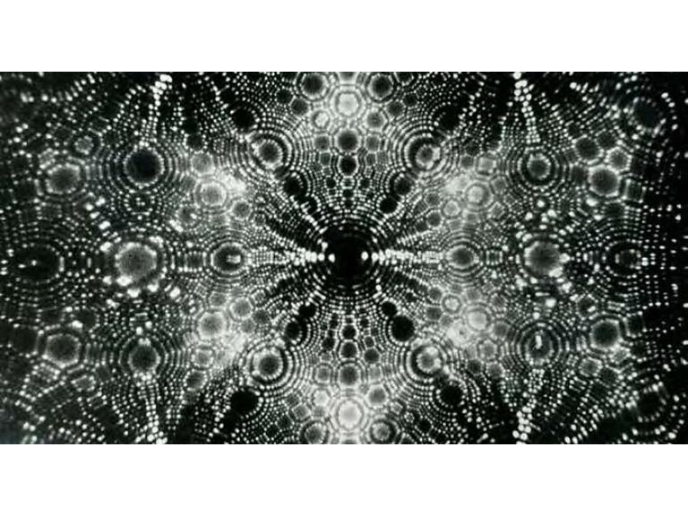
How the Field Ion Microscope Works
The field ion microscope did not produce traditional photographs of atoms. Instead, it used a sharp tungsten tip placed in an ultra-high vacuum chamber filled with helium gas. The tip was cooled with liquid nitrogen, and a positive voltage was applied, causing tungsten ions to be repelled. These ions were collected on a phosphor screen, creating a magnified image at atomic resolution.
“Nowadays, to be able to ‘see’ atoms is remembered as a major achievement in the field of microscopy,” said Mauricio Terrones, head of the Department of Physics at Penn State.
Impact and Advancements in Microscopy
Müller’s work sparked a revolution in resolution, influencing the development of atomic resolution imaging techniques. Since 1955, these techniques have advanced significantly, allowing scientists to visualize individual atoms, reveal crystal structures, and determine atomic bonding and elemental compositions.
Students of atomic imaging, such as those studying atom probe tomography (APT), often begin with Müller’s inventions. Oscar Lopez, a former postdoctoral researcher, emphasized Müller’s lasting impact: “People in the field have a real respect for Müller and his legacy.”
Modern Applications and Innovations
Today, Müller’s contributions are evident in research at Penn State and beyond. Raymond Schaak, a professor of materials chemistry, explained how his research utilizes atomic resolution imaging to improve catalytic reactions in clean energy and solar cells. “Our chemistry research works to place atoms in precise locations within nanostructured materials,” he noted.
Danielle Reifsnyder Hickey, an assistant professor, highlighted the influence of Müller’s work on modern technology. “Using aberration-corrected transmission electron microscopy, my lab contributes to creating powerful new technologies,” she said, referencing advancements in electronics like smartphones and computers.
The Future of Atomic Resolution Imaging
Penn State’s Materials Research Institute, home to the Materials Characterization Laboratory, continues to push the boundaries of atomic and nanoscale resolution imaging. Researchers from various disciplines, including chemistry and physics, are building on Müller’s legacy to explore new possibilities.
The implications of Müller’s work extend far beyond his time, inspiring technologies that were once unimaginable. As atomic resolution imaging continues to evolve, its applications in science and industry promise to drive innovation for decades to come.