


In a groundbreaking study, scientists have identified a rare group of blue proteins known as cryorhodopsins, found exclusively in cold environments such as glaciers and high mountains. These proteins, derived from cold-adapted microbes, have the potential to revolutionize the design of molecular on-off switches for cellular activity, offering promising applications in science and medicine.
Rhodopsins, akin to chlorophyll, are proteins that enable organisms to absorb sunlight for energy. Some rhodopsins can be engineered to function as light-operated power switches for electrical activity in cells. The recent discovery of cryorhodopsins, which absorb red light—a rare property—opens new avenues for scientific exploration.
Unveiling the Secrets of Cryorhodopsins
Kirill Kovalev, a structural biologist and EIPOD Postdoctoral Fellow at EMBL Hamburg and EMBL-EBI, has been captivated by rhodopsins for years. His research focuses on understanding the unique features of these proteins, which could have untapped potential. “In my work, I search for unusual rhodopsins and try to understand what they do,” Kovalev explained. “Such molecules could have undiscovered functions that we could benefit from.”
While browsing protein databases, Kovalev stumbled upon an unusual feature common to microbial rhodopsins found in cold environments. These cryorhodopsins, despite evolving thousands of kilometers apart, were strikingly similar, suggesting their essential role in surviving harsh conditions. Kovalev named them ‘cryorhodopsins’ to reflect their cold habitat.
Color and Functionality
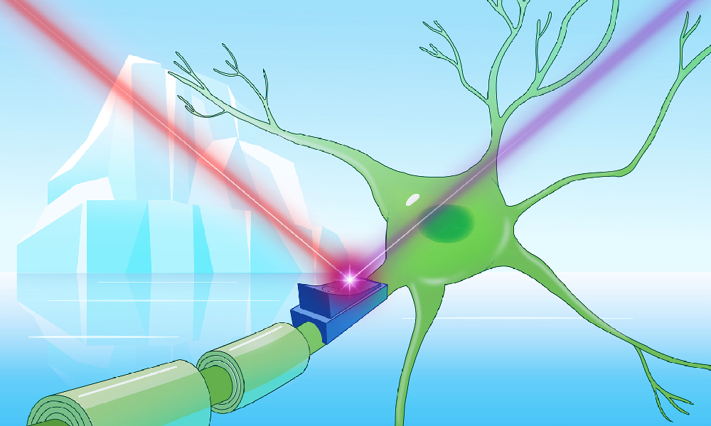
The color of rhodopsins is a crucial characteristic, as it determines the wavelengths of light they absorb and reflect. Most rhodopsins are pink-orange, absorbing green and blue light. However, Kovalev’s lab revealed an unexpected diversity of colors among cryorhodopsins, with some exhibiting the sought-after blue hue. This discovery is significant because blue rhodopsins, activated by red light, penetrate tissues more deeply and non-invasively.
“I can actually tell what’s going on with cryorhodopsin simply by looking at its color,” laughed Kovalev.
By applying advanced structural biology techniques, Kovalev identified the structural feature responsible for the blue color. “Now that we understand what makes them blue, we can design synthetic blue rhodopsins tailored to different applications,” he noted.
Potential Applications and Future Directions
Subsequent experiments demonstrated cryorhodopsins’ potential as optogenetic tools. When exposed to UV light, cryorhodopsins induced electric currents in cultured brain cells. The cells’ excitability varied depending on the light color used, highlighting their potential for research, biotechnology, and medical applications.
“New optogenetic tools to efficiently switch the cell’s electric activity both ‘on’ and ‘off’ would be incredibly useful,” said Tobias Moser, Group Leader at the University Medical Center Göttingen.
While cryorhodopsins are not yet ready for practical use, they serve as promising prototypes. Kovalev emphasized, “They have all the key features that, based on our findings, could be engineered to become more effective for optogenetics.”
Evolutionary Insights
Further investigations revealed that cryorhodopsins might act as photosensors, allowing microbes to detect UV light—a property previously unheard of among rhodopsins. This ability could help microbes in cold environments, such as mountain tops, to sense and protect themselves from harmful UV radiation.
“We suspect that cryorhodopsins evolved their unique features not because of the cold, but to let microbes sense UV light, which can be harmful to them,” Kovalev suggested.
Collaborative efforts using AI tools like AlphaFold provided insights into the potential mechanisms by which cryorhodopsins transmit light-sensitive signals within cells. These findings hint at a broader range of functions for associated proteins, even in organisms lacking cryorhodopsins.
Overcoming Technical Challenges
Studying cryorhodopsins required overcoming significant technical hurdles. Their nearly identical structures meant that even slight atomic changes could alter their properties. Kovalev utilized a 4D structural biology approach, combining X-ray crystallography and cryo-electron microscopy, to explore these molecules in detail.
“I chose to do my postdoc at EMBL Hamburg because of the unique beamline setup that made my project possible,” Kovalev remarked, expressing gratitude for the support received from the P14 beamline team.
Additionally, the extreme light sensitivity of cryorhodopsins necessitated working with samples in near darkness, posing further challenges for Kovalev’s collaborators.
Building Skills and Collaborations
Kovalev’s postdoctoral journey in the EIPOD4 fellowship program has been transformative, allowing him to engage in ambitious research projects with multiple EMBL group leaders. His collaborative efforts extended across institutions in Germany, the Netherlands, and beyond.
“During my EIPOD fellowship, I progressed from conducting independent research to engaging in close collaborations with leading scientists worldwide,” Kovalev reflected. “This hands-on learning experience has been truly invaluable.”
As the study of cryorhodopsins continues, their unique properties promise to unlock new scientific frontiers, offering insights into both the natural world and potential technological applications.