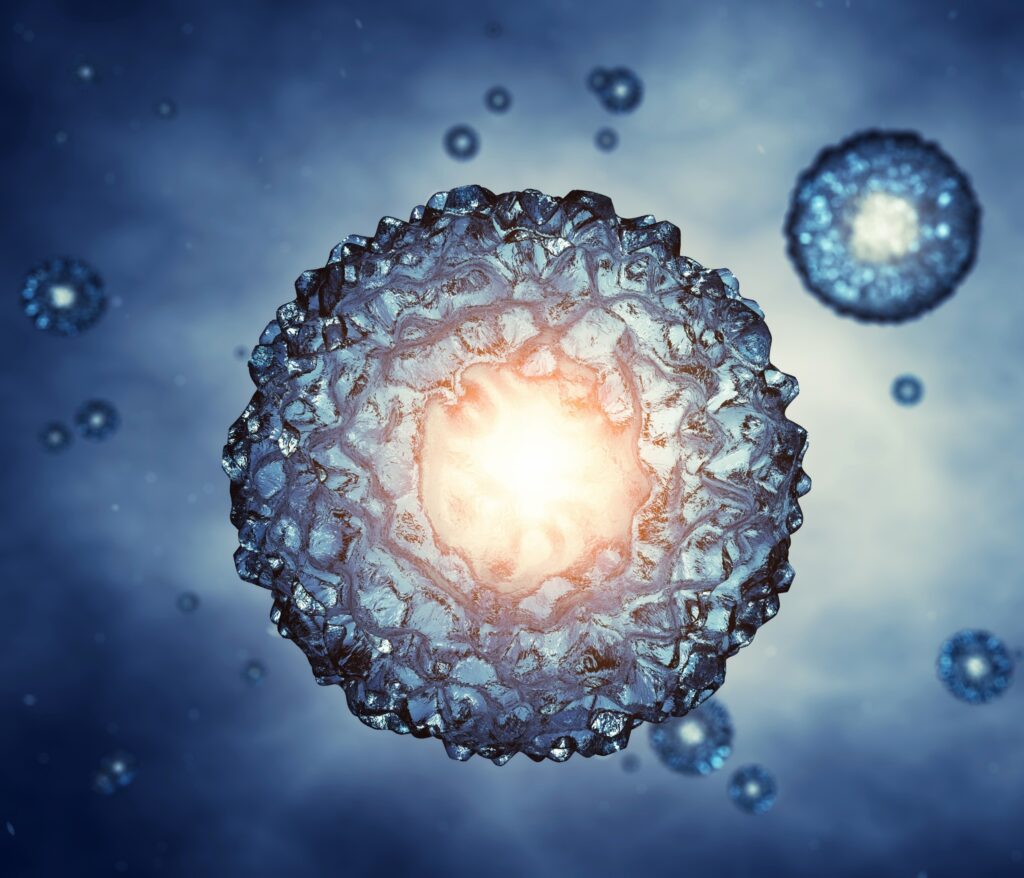
Every human life begins as a cluster of cells that soon face critical decisions. Within weeks, these cells must choose their fates—some forming the nervous system, others the gut, heart, or muscle. For decades, scientists have understood the broad strokes of this developmental story but lacked the intricate details. Much of the information came from mouse studies, which often proved misleading when applied to humans.
A groundbreaking study from the University of Cambridge has changed this narrative. Researchers have successfully mapped human embryos between days 7 and 21 with single-cell precision, capturing the pivotal moments when pluripotent cells transition from having open potential to adopting organized roles. The result is a comprehensive reference atlas that illuminates how early human development unfolds, moving beyond theoretical models.
Critical Days of Development
The days following implantation are crucial in defining the course of development. During this period, epiblast cells spread out and fold into distinct layers known as germ layers: ectoderm, mesoderm, and endoderm. These layers serve as the template for all body systems. Timing is vital here, as humans extend these events over longer periods than mice, allowing signals more time to fine-tune.
For years, scientists have pieced together this developmental puzzle from fragmented data. Now, with embryos analyzed on a daily basis, the sequence of events appears much clearer. The atlas not only shows when cells make critical decisions but also reveals how their location within the embryo influences those decisions.
Mapping Embryo Cell Roles
The research team employed single-cell RNA sequencing to capture the activities of individual cells. Mesoderm precursors begin activating muscle and bone programs, while endoderm cells initiate digestive and liver gene expression. Ectoderm cells start setting up the nervous system. Each developmental path unfolds according to its own timing, yet none occur in isolation.
Spatial maps added another dimension to the study. Cells do not express genes randomly; their position within the embryo is significant. The data reveal that cell identity evolves in sync with physical context, as if a silent conductor orchestrates the process.
Limits of Lab Models
Stem cell models, such as gastruloids and blastoids, have provided scientists with glimpses into development. However, how accurately do these models reflect reality? While models mimic the broad picture, they often miss finer transitions. Germ layers emerge, but their boundaries are blurred, and some cell types never materialize.
This new atlas offers researchers a benchmark to measure accuracy. By comparing lab-grown systems with real embryos, discrepancies become apparent. The findings are not discouraging but rather instructive, allowing models to be adjusted, refined, and tested against an authentic reference.
Lessons from Evolution
The study extends beyond human development by comparing expression patterns across mammals. Core signals such as WNT and NODAL echo across species, yet humans slow the developmental tempo, prolonging early stages. This pause creates differences that ripple through the entire development process.
This insight helps explain why mouse study results often falter when translated to humans. Evolution has conserved the toolkit but not the schedule. By mapping humans directly, the study highlights what makes our species unique and what remains shared with others.
Why the Embryo Map Matters
Reproductive medicine stands to benefit significantly from this research, as it provides precise timelines for when embryos pass vital milestones. IVF specialists, for example, can use these insights to better assess embryo health. Regenerative medicine also gains direction, as stem cells can be more reliably guided toward organ-specific paths when early signals are mapped in detail.
Organoid research will also benefit. As scientists grow mini-organs in lab settings, they can now compare them against natural blueprints, increasing confidence that what forms in the lab resembles what forms in life.
Walking the Ethical Line
Embryo research often faces ethical scrutiny. The study’s ethical statement emphasizes that all work adhered to strict guidelines, including the 14-day limit. Embryos were donated with consent, and experiments followed international standards.
As stem cell models become more realistic, ethical debates will intensify. Are models embryos in spirit, even if not in law? The paper does not shy away from this tension, framing progress within responsibility and reminding readers that science must advance without losing sight of ethical boundaries.
Future of the Embryo Map
The atlas does more than answer questions—it sets a foundation for future research. Scientists now have a benchmark to guide work worldwide, from IVF to organoid design, offering both detail and direction. Perhaps the most significant shift is in perspective. For the first time, researchers can observe early human development at single-cell resolution, tracing how potential narrows into destiny and how minute shifts create the structure of life.
This study captures a rare view: the earliest choreography of human existence. Cells do not wander randomly; they follow cues, take positions, and form a body plan that will last a lifetime. With this atlas, we see not only how humans grow but also why we differ from other animals and where models need correction.
Science now has a sharper lens on the dawn of life, and the responsibility is to use it wisely for the future of human health. The study is published in the journal Nature.
—
Like what you read? Subscribe to our newsletter for engaging articles, exclusive content, and the latest updates.
Check us out on EarthSnap, a free app brought to you by Eric Ralls and Earth.com.


