


Red blood cells play a crucial role in transporting oxygen and supporting immune functions in the human body. However, when these cells become abnormally shaped, they can signal serious health conditions such as diabetes, malaria, hereditary blood disorders, and vascular diseases. Traditionally, diagnosing these abnormalities has relied heavily on optical microscopy, a method that involves fluorescent staining and manual inspection by trained experts. This approach, while widely used, is fraught with limitations: it is time-consuming, subjective, and often misses subtle changes in individual cells.
In a groundbreaking development, a research team led by Prof. Nan Zeng at Tsinghua Shenzhen International Graduate School has introduced a novel method using polarized light technology. This new technique, known as dual-angle Mueller matrix polarimetry (DMMP), offers a label-free solution to analyze red blood cell deformations at the single-cell level.
Revolutionizing Blood Cell Analysis
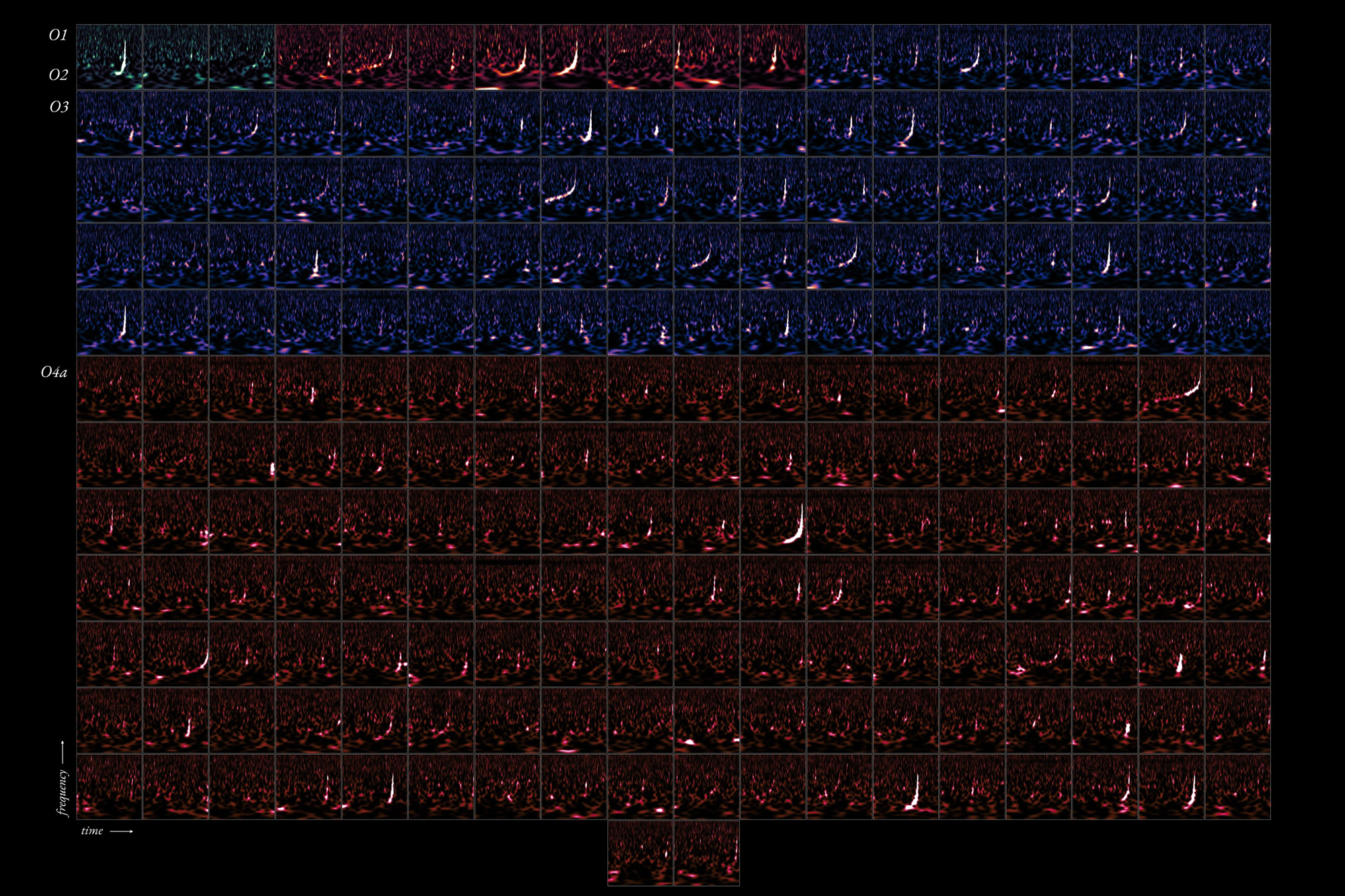
The DMMP method stands out by utilizing polarized light to examine individual blood cells, measuring how they alter the polarization state of light. This innovative system shines polarized light on cells and captures their unique polarization signatures. By integrating theoretical modeling with machine learning algorithms, the researchers have identified six specific polarization feature parameters. These parameters quantitatively describe cell size, shape, refractive index, and surface characteristics.
To validate their approach, the team tested red blood cells under various stress conditions that mimic disease states. The results were promising, showing that the polarization parameters could effectively distinguish between normal cells and those with abnormal deformations. Furthermore, when analyzing mixed blood samples containing both healthy and abnormal cells, a Random Forest classifier using these parameters achieved over 94% accuracy in determining cell type proportions.
Advantages Over Traditional Methods
The DMMP technology offers several significant advantages over traditional methods. It eliminates the need for staining or complex sample preparation, allowing for the analysis of hundreds of cells per minute, which is ideal for high-throughput screening. Moreover, it provides objective, quantitative measurements rather than subjective visual assessments, potentially leading to more accurate diagnoses.
This label-free approach could be readily adapted for routine blood screenings in hospitals and clinics, enabling earlier disease detection, more accurate diagnosis, and better monitoring of treatment responses for blood-related disorders. The implications of this technology are vast, offering a new frontier in medical diagnostics.
Implications and Future Prospects
The introduction of DMMP technology represents a significant leap forward in medical diagnostics. By providing a rapid, objective, and cost-effective method for analyzing red blood cells, this tool could revolutionize the way blood-related disorders are detected and monitored. The potential for earlier disease detection and improved patient outcomes is substantial.
As Prof. Nan Zeng’s team continues to refine and expand the capabilities of this technology, the medical community is optimistic about its applications. The work, titled “Analysis of erythrocyte deformation characteristics based on dual-angle Mueller matrix measurement,” was published in Frontiers of Optoelectronics on December 3, 2025, marking a pivotal moment in the field of optoelectronics and medical diagnostics.
Looking ahead, the integration of DMMP technology into clinical settings could transform routine blood screenings, offering a more efficient and accurate method for diagnosing and monitoring a range of health conditions. As research progresses, the potential for this technology to impact global health is immense, paving the way for a new era in medical diagnostics.