


Successful cancer surgery hinges on a surgeon’s ability to excise tumors while preserving healthy tissue. Currently, surgeons employ fluorescent dyes to distinguish cancer cells from healthy ones, but these dyes are not infallible, often illuminating healthy tissues as well. In a groundbreaking development, researchers from the University of Tokyo have introduced a bioorthogonal fluorescence probe paired with a specialized reporter enzyme, capable of selectively activating the probe at tumor sites. This innovation enables high-contrast tumor visualization with minimal background interference, though the study has so far only been conducted on mice.
Cancer remains a global health challenge, affecting millions worldwide. Many patients opt for surgery, hoping for complete tumor removal without affecting surrounding healthy tissues. Over the years, various tools and techniques have been devised to enhance surgical outcomes, with visual imaging methods like fluorescent dyes proving invaluable. However, a significant drawback is that some probes can be inadvertently activated by endogenous enzymes in healthy tissues, creating background fluorescence and complicating surgical decisions. Conversely, some cancer cells may remain unmarked, increasing the risk of recurrence.
Innovative Approach to Tumor Imaging
Associate Professor Ryosuke Kojima from the Laboratory of Chemical Biology and Molecular Imaging at the University of Tokyo explains, “Our group acknowledged this current shortcoming and improved upon this way to make cancer cells light up inside the body. In tests on mice, we delivered a special enzyme to tumors and used a fluorescence probe that only turns on when that enzyme is present.”
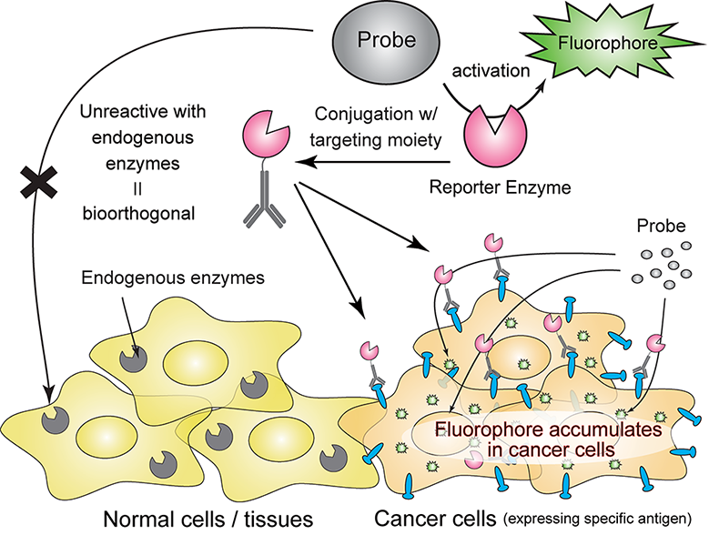
“Older probes often light up healthy tissue by mistake, creating background noise, but our highly selective, or bioorthogonal, dye probe is designed to stay completely off unless it meets its matching engineered enzyme,” Kojima added.
Through a process of directed evolution involving repeated mutation and selection, Kojima and his team developed an enzyme capable of strongly activating the probe within living animals. This novel probe is resistant to activation by natural enzymes in the body, reducing unwanted background glow. When tested on mice with peritoneal cancer, the engineered enzyme successfully reached the tumors, and the probe illuminated them as expected.
Potential for Broader Applications
This development could revolutionize surgical oncology by enabling the visualization of tiny, millimeter-sized tumor lesions with extremely low background noise. “This allowed us to see tiny, millimeter-sized tumor lesions with extremely low background noise, a level of contrast that could be very useful during surgery,” said Kojima.
In the short term, this system could serve as a powerful research tool, and in the long term, it may assist surgeons in more thoroughly removing tumors by clearly highlighting cancer cells. However, a significant challenge for clinical application will be ensuring that the engineered enzyme does not provoke an unwanted immune response in patients.
The system’s adaptability to other cancer types is another promising aspect. Many cancers present specific antigens, or markers of tumor tissue, which could be targeted by swapping the tumor-targeting component. This flexibility means the same enzyme-probe pair could potentially be redirected to other cancer types.
Future Implications and Challenges
Looking further ahead, this research could pave the way for highly targeted drug delivery. Instead of fluorescent dyes, cancer-fighting drugs could be delivered precisely to the required sites. However, as Kojima cautions, “It’s still early days, the trials have only been done in mice, and much work is needed before it’s deemed safe enough for human trials.”
The announcement comes at a time when precision medicine is gaining momentum, offering hope for more effective and less invasive cancer treatments. As researchers continue to refine this technology, the potential for improved surgical outcomes and targeted therapies represents a significant step forward in the fight against cancer.