


Scientists at the Center for Cell Dynamics, School of Biological and Behavioral Sciences, Queen Mary University of London, in collaboration with Carl Zeiss, have unveiled a groundbreaking live-cell imaging technique. This innovative method combines an exceptional resolution of 60 nanometers with fluorescence recovery after photobleaching, significantly reducing light-induced cellular damage. The development promises to revolutionize the study of intricate cellular processes by offering unprecedented clarity, thus opening new avenues for understanding fundamental biological mechanisms such as DNA repair and chromosome dynamics.
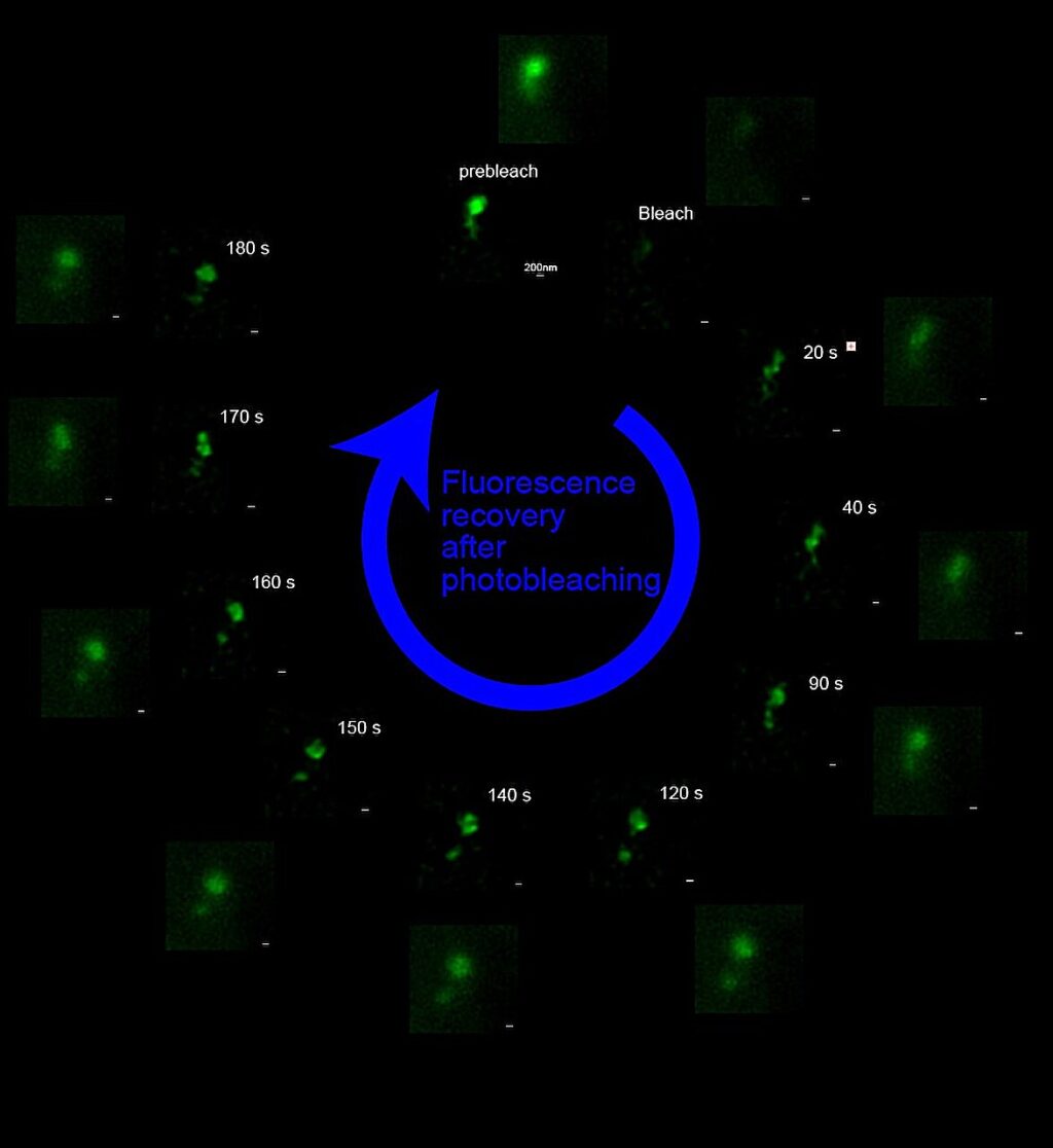
The team, led by Professor Viji Draviam, has ingeniously combined Lattice Structured Illumination Microscopy (diSIM/SIM²) with Fluorescence Recovery After Photobleaching (FRAP) to create a novel method termed FRAP-SR (FRAP in Super-Resolution regime). This technique overcomes the limitations of traditional microscopy and earlier super-resolution methods, which often suffer from phototoxicity, hindering the study of delicate biological events in living cells. The findings are published on the bioRxiv preprint server.
Innovative Approach to Live-Cell Imaging
“Our FRAP-SR approach enables us to visualize structures as small as 60 nanometers within living cells—a scale previously inaccessible for dynamic studies without causing significant cellular stress,” explains Professor Draviam. “This resolution, 2000 times smaller than the width of a human hair, allows us to probe the nanoscale organization and behavior of cellular components in real-time.”
Using FRAP-SR, the researchers investigated the dynamics of 53BP1, a key protein involved in the repair of double-strand DNA breaks. Their high-resolution live-cell imaging revealed that 53BP1 forms liquid-like condensates with surprising complexity. Some of these foci appeared as stable, compact structures, while others exhibited more fluid, dynamic shapes.
Full nuclei with DNA damage spots decorated by green cloud-like 53BP1-foci.
Unveiling Subcompartments in Cellular Structures
By applying FRAP-SR, the team discovered that the amorphous 53BP1 foci contain distinct subcompartments with varying protein mobility, suggesting functional specialization within these repair centers. In contrast, compact foci displayed uniform recovery after photobleaching but showed greater heterogeneity in recovery rates between different foci. The study also revealed that the dynamics of these foci are influenced by cellular conditions, such as recovery from DNA replication stress.
Professor Draviam highlights the potential of this innovation: “FRAP-SR provides a powerful tool to dissect the dynamic architecture of protein assemblies at the nanoscale in living cells. It allows us to investigate fundamental cellular processes, particularly those sensitive to light exposure, with unprecedented detail and minimal perturbation. This will transform the field of optogenetics in the super-resolution regime. It will also enable the development of new anti-cancer drugs that target DNA damage repair pathways that are dynamic.”
Implications for Future Research and Medicine
This advancement holds significant promise for cell biology researchers studying a wide range of light-sensitive processes, including DNA damage response, chromosome organization, mitochondrial dynamics, and cellular senescence. The ability to study these processes at high resolution in living cells without inducing damage will undoubtedly accelerate discoveries in these fields.
The global DNA repair drugs market was valued at approximately USD 9.18 billion in 2024 and is projected to reach USD 13.97 billion by 2030, growing at a compound annual growth rate (CAGR) of around 7.2%.
The team has shown how the DNA damage marker 53BP1 can be exploited in live-cells using FRAP-SR to accelerate the development of novel DNA repair drugs or drug candidates relevant to personalized medicine.
Technological Collaboration and Future Prospects
The ZEISS Elyra 7 system, enhanced with FRAP capabilities from Rapp OptoElectronics, was instrumental in this study, providing the advanced super-resolution imaging necessary to resolve the subcompartments of 53BP1 foci for the first time. Professor Draviam’s collaboration with Zeiss and Rapp OptoElectronics to integrate FRAP and structured illumination microscopy allowed for precise quantification of protein dynamics.
As the scientific community continues to explore the potential of FRAP-SR, the implications for both fundamental research and clinical applications are vast. This breakthrough not only enhances our understanding of cellular processes but also paves the way for the development of targeted therapies in personalized medicine, particularly in the realm of cancer treatment.
More information: Chengchen Wu et al, FRAP-in-SR: Fluorescence recovery in the Super-Resolution regime reveals subcompartments of 53BP1 foci, bioRxiv (2025). DOI: 10.1101/2025.05.07.652606 Journal information: bioRxiv