


In a groundbreaking advancement for both research and medical applications, a team of scientists and engineers at the Massachusetts Institute of Technology (MIT) has developed a new microscope system that can peer deep into brain tissues to observe molecular activity at the single-cell level. This innovative technology, which utilizes sound to achieve unprecedented imaging depth, offers new insights into brain activity in regions like the hippocampus, which have long been challenging to study.
The study, published in the journal Light: Science and Applications, showcases the system’s capability to detect NAD(P)H, a molecule closely associated with cell metabolism and neuronal electrical activity. The team successfully demonstrated this by imaging through samples like a 1.1-millimeter “cerebral organoid” and a 0.7-millimeter-thick slice of mouse brain tissue. According to neuroscientist Mriganka Sur, a corresponding author of the study, “The major advance here is to enable us to image deeper at single-cell resolution.”
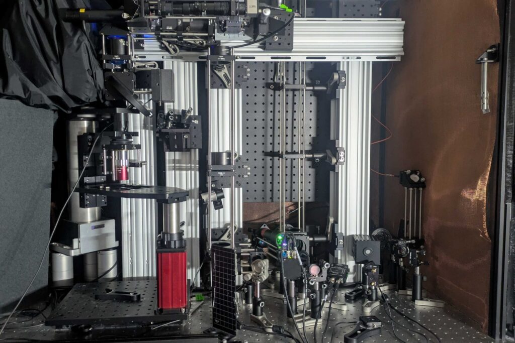
Revolutionary Imaging Techniques
The new system achieves its remarkable depth and clarity by combining several cutting-edge technologies. It employs a method known as “three-photon” excitation, which uses an intense, extremely short burst of light at three times the normal absorption wavelength. This approach allows the light to penetrate deeper into brain tissue with less scattering, similar to how fog lamps work. The resulting weak fluorescent signal from NAD(P)H is complemented by sound waves generated from thermal expansion within the cell, detected by an ultrasound microphone.
Co-lead author and mechanical engineering postdoc W. David Lee, who conceived the microscope’s design, explains, “We merged all these techniques – three-photon, label-free, photoacoustic detection – into one process to establish this ‘Multiphoton-In and Acoustic-Out’ platform.” The system’s ability to produce high-resolution images without external labels or genetically engineered fluorescence marks a significant leap forward in neuroscience research.
Potential Applications in Neuroscience and Medicine
The implications of this breakthrough are vast, particularly in the fields of neuroscience and clinical medicine. The technology’s label-free nature means it could be safely used in humans, potentially during brain surgeries. NAD(P)H levels are known to vary in conditions such as Alzheimer’s disease, Rett syndrome, and seizures, making it a valuable biomarker for these and other neurological disorders.
Lee, who has previously demonstrated the utility of NAD(P)H imaging in wound care through his company Precision Healing, Inc., sees great potential for this technology in diagnosing and understanding brain disorders. “In principle, it should work,” he says, expressing confidence in the system’s future applications.
Next Steps and Future Research
The next challenge for the MIT team is to demonstrate the system’s capabilities in a living animal, moving beyond in vitro and ex-vivo tissues. This requires technical adjustments, such as positioning the microphone on the same side as the light source. Lee anticipates that full imaging at depths of 2 millimeters in live brains is achievable, given the promising results of the current study.
Funding for this research was provided by various sources, including the National Institutes of Health, the Simon Center for the Social Brain, the lab of Peter So, The Picower Institute for Learning and Memory, and the Freedom Together Foundation. As the team continues to refine their techniques, the potential for this technology to transform our understanding of brain function and disease remains immense.
With continued development, this innovative imaging system could soon become a staple tool in neuroscience research and clinical practice, offering unprecedented insights into the intricate workings of the human brain.