


In a groundbreaking study, researchers at the Flaum Eye Institute and Del Monte Institute for Neuroscience at the University of Rochester have unveiled a unique response mechanism of the retina to injury. Unlike other tissues in the body, where neutrophils—immune cells found in the blood—are the first responders to infections or injuries, the retina relies on microglia, the brain’s immune cells, to address damage to photoreceptor cells. This discovery could significantly impact the understanding and treatment of vision loss affecting millions of Americans.
“This finding has high implications for what happens for millions of Americans who suffer vision loss through loss of photoreceptors,” stated Jesse Schallek, PhD, associate professor of Ophthalmology and senior author of the study published in eLife. “This association between two key immune cell populations is essential knowledge as we build new therapies that must understand the nuance of immune cell interactions.”
Understanding the Retina’s Unique Immune Response
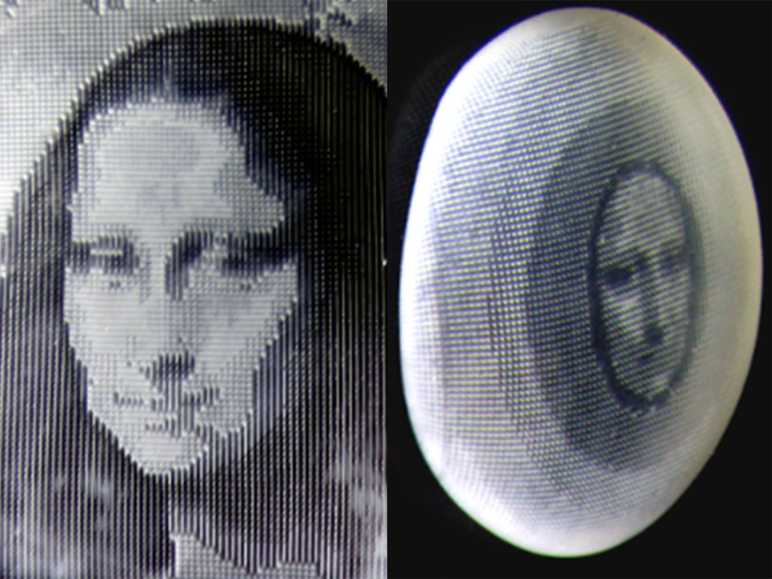
Using adaptive optics imaging, a sophisticated camera technology developed by the University of Rochester, researchers studied the retinas of mice with photoreceptor damage. This technology allows for the imaging of single neurons and immune cells inside the living eye, providing unprecedented insight into cellular interactions. The study revealed that while both neutrophil and microglia cells are present in the retina, only microglia respond to photoreceptor injury. Notably, they do not recruit neutrophils to assist in the repair process.
“What is remarkable here is that the passing neutrophils are so close to the reactive microglia, and yet they do not signal to them to assist in damage recovery,” said Schallek. “This is notably different than what is seen in other areas of the body where neutrophils are the first to respond to local damage and mount an early and robust response.”
Implications for Vision Health
Photoreceptor cells, unique to the retina, are responsible for converting light into electrical and chemical signals, which are then communicated to the brain to enable vision. Diseases such as age-related macular degeneration, retinitis pigmentosa, and cone-rod dystrophy can damage and kill these cells, leading to vision loss. Currently, there is no cure for these conditions, making this research particularly significant.
The study suggests that a form of cloaking occurs during retinal injury, potentially protecting the retina from an influx of immune cells that could exacerbate damage. This discovery opens new avenues for developing therapies that could better manage or even prevent vision loss by leveraging the retina’s natural immune response.
Expert Insights and Future Directions
The research, led by first author Derek Power, a laboratory technician in the Schallek lab, and co-authored by Justin Elstrott, PhD, of Genentech Inc., highlights the importance of understanding immune cell interactions in developing effective treatments. Supported by the National Eye Institute, Research to Prevent Blindness, the Dana Foundation, and a Collaborative Research Grant from Genentech, Inc., this study is a pivotal step in vision science.
Looking ahead, researchers are keen to explore how these findings can be translated into clinical applications. By visualizing the dynamics of single cells as they communicate, scientists hope to develop targeted therapies that harness the retina’s unique immune response, potentially offering new hope for those affected by degenerative eye diseases.
As the study progresses, the scientific community remains optimistic about the potential breakthroughs in understanding and treating vision loss. The continued collaboration between research institutions and industry partners will be crucial in advancing these efforts and ultimately improving outcomes for individuals with vision impairments.