


The signals that drive many of the brain and body’s most essential functions—consciousness, sleep, breathing, heart rate, and motion—course through bundles of “white matter” fibers in the brainstem. However, imaging systems have struggled to finely resolve these crucial neural cables, leaving researchers and doctors with limited capability to assess how they are affected by trauma or neurodegeneration.
In a groundbreaking study, a team of researchers from MIT, Harvard University, and Massachusetts General Hospital has unveiled an AI-powered software capable of automatically segmenting eight distinct bundles in any diffusion MRI sequence. This new tool, known as the BrainStem Bundle Tool (BSBT), has been made publicly available and is detailed in an open-access study published on February 6 in the Proceedings of the National Academy of Sciences.
Advancements in Brain Imaging
The research, led by MIT graduate student Mark Olchanyi, reveals that BSBT can identify distinct patterns of structural changes in patients with Parkinson’s disease, multiple sclerosis, traumatic brain injury, and even shed light on Alzheimer’s disease. Remarkably, BSBT has also been used retrospectively to track bundle healing in a coma patient, reflecting the patient’s seven-month recovery journey.
“The brainstem is a region of the brain that is essentially not explored because it is tough to image,” says Olchanyi, a doctoral candidate in MIT’s Medical Engineering and Medical Physics Program. “People don’t really understand its makeup from an imaging perspective. We need to understand what the organization of the white matter is in humans and how this organization breaks down in certain disorders.”
Professor Emery N. Brown, Olchanyi’s thesis supervisor and co-senior author of the study, adds, “The brainstem is one of the body’s most important control centers. Mark’s algorithms are a significant contribution to imaging research and to our ability to understand the regulation of fundamental physiology.”
Building the Algorithm
Diffusion MRI is a technique that helps trace the long branches, or “axons,” that neurons extend to communicate with each other. These axons are typically clad in a sheath of fat called myelin, and the diffusion of water along these axons is a key focus of diffusion MRI. However, segmenting the distinct bundles of axons in the brainstem has been challenging due to their small size and the masking effects of brain fluids and bodily motions.
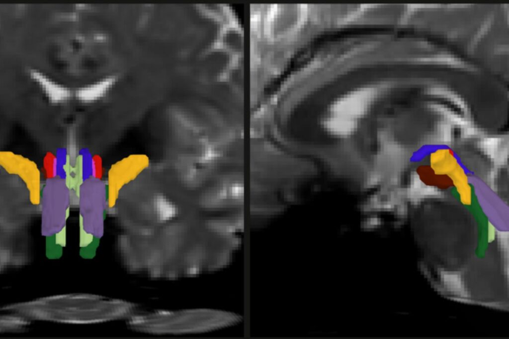
Olchanyi’s work aimed to overcome these obstacles by developing an AI algorithm. BSBT works by tracing fiber bundles that enter the brainstem from neighboring brain areas, producing a “probabilistic fiber map.” A convolutional neural network then combines this map with multiple imaging channels to distinguish eight individual bundles.
To train the neural network, Olchanyi used 30 live diffusion MRI scans from volunteers in the Human Connectome Project, manually annotating them to teach the network how to identify the bundles. The tool’s accuracy was validated against dissections of post-mortem human brains.
Potential Novel Biomarkers
Once trained and validated, the research team tested whether the ability to segment distinct fiber bundles could track variations in bundle volume and structure due to disease or injury, potentially creating novel biomarkers. Despite the difficulty in examining the brainstem, many studies indicate that neurodegenerative diseases often affect this region early in their progression.
Olchanyi and his colleagues applied BSBT to datasets of diffusion MRI scans from patients with Alzheimer’s, Parkinson’s, MS, and traumatic brain injury. The tool measured bundle volume and “fractional anisotropy” (FA), a proxy for white matter structural integrity. The results showed consistent patterns of changes in the bundles across different conditions.
BSBT proved more accurate than other classifier methods in discriminating between patients with health conditions versus controls. The authors noted, “BSBT can be a key adjunct that aids current diagnostic imaging methods by providing a fine-grained assessment of brainstem white matter structure and, in some cases, longitudinal information.”
Implications and Future Directions
The study also highlighted BSBT’s potential in prognostics. In the case of a 29-year-old man with severe traumatic brain injury, BSBT was applied to scans taken during his seven-month coma. The tool showed that the man’s brainstem bundles had been displaced but not cut, and as they healed, the bundles moved back into place.
The authors concluded that BSBT “has substantial prognostic potential by identifying preserved brainstem bundles that can facilitate coma recovery.”
With funding from various prestigious institutions, including the National Institutes of Health and the U.S. Department of Defense, this study marks a significant advancement in neuroscience research. The potential applications of BSBT in clinical settings could transform how neurodegenerative diseases and brain injuries are diagnosed and monitored, offering new hope for patients and healthcare providers alike.