


A groundbreaking artificial intelligence (AI) tool has been developed to significantly ease and reduce the cost of training medical imaging software, even when only a limited number of patient scans are available. This innovative tool enhances medical image segmentation—a process where each pixel in an image is labeled according to what it represents, such as cancerous or normal tissue—traditionally performed by highly trained experts.
The announcement comes as deep learning methods, which have shown promise in automating this labor-intensive task, face the challenge of being heavily data-dependent. According to Li Zhang, a Ph.D. student in the Department of Electrical and Computer Engineering at the University of California San Diego, these methods require extensive datasets of pixel-by-pixel annotated images, which are costly and time-consuming to create. This poses a significant hurdle for many medical conditions and clinical settings where such data is scarce.
Innovative Approach to Data Scarcity
In response to these challenges, Zhang and a team of researchers led by UC San Diego professor Pengtao Xie have developed an AI tool capable of learning image segmentation from a minimal number of expert-labeled samples. This tool reduces the data requirement by up to 20 times, potentially leading to faster and more affordable diagnostic tools, particularly in resource-limited hospitals and clinics.
Their findings, published in Nature Communications, highlight the potential of this tool to transform medical diagnostics. “This project was born from the need to break this bottleneck and make powerful segmentation tools more practical and accessible, especially for scenarios where data are scarce,” Zhang, the study’s first author, explained.
Testing and Performance
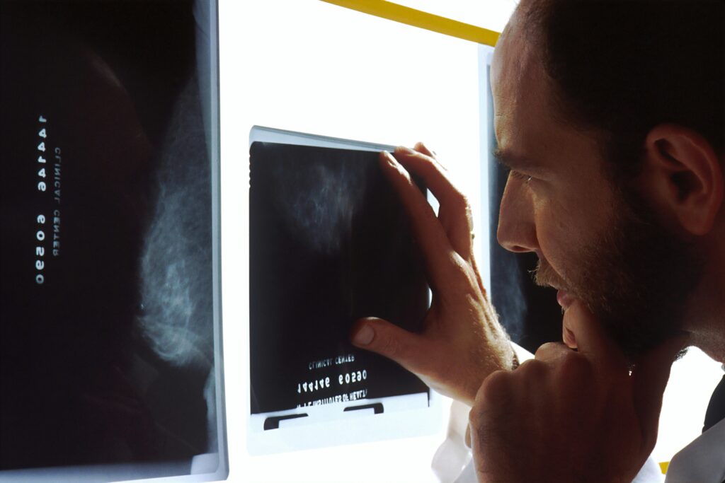
The AI tool underwent rigorous testing across various medical image segmentation tasks. It successfully identified skin lesions in dermoscopy images, breast cancer in ultrasound scans, placental vessels in fetoscopic images, polyps in colonoscopy images, and foot ulcers in standard camera photos. The method was even extended to 3D images, such as those used to map the hippocampus or liver.
In settings with extremely limited annotated data, the AI tool improved model performance by 10 to 20% compared to existing approaches, requiring 8 to 20 times less real-world training data while often matching or outperforming them.
Zhang illustrated how this AI tool could assist dermatologists in diagnosing skin cancer. Instead of needing to gather and label thousands of images, a trained expert might only need to annotate 40. The AI tool could then use this small dataset to identify suspicious lesions from a patient’s dermoscopy images in real-time, thus aiding doctors in making faster, more accurate diagnoses.
Mechanics of the AI System
The system operates in stages. Initially, it learns to generate synthetic images from segmentation masks—color-coded overlays that indicate which parts of an image are healthy or diseased. It then creates new, artificial image-mask pairs to augment a small dataset of real examples. A segmentation model is trained using both sets of data. Through a continuous feedback loop, the system refines the images it creates based on their effectiveness in improving the model’s learning.
“Rather than treating data generation and segmentation model training as two separate tasks, this system is the first to integrate them together. The segmentation performance itself guides the data generation process,” Zhang noted. “This ensures that the synthetic data are not just realistic, but also specifically tailored to improve the model’s segmentation capabilities.”
Future Prospects and Implications
Looking ahead, the research team plans to enhance the AI tool’s intelligence and versatility. They aim to incorporate direct feedback from clinicians into the training process, ensuring the generated data is more applicable to real-world medical scenarios.
The development of this AI tool represents a significant advancement in medical imaging technology, offering the potential to democratize access to high-quality diagnostic tools worldwide. The work was supported by the National Science Foundation (IIS2405974 and IIS2339216) and the National Institutes of Health (R35GM157217 and R21GM154171).
As the healthcare industry continues to embrace AI, tools like this could play a pivotal role in overcoming data limitations, ultimately leading to more efficient and accessible healthcare solutions.