


If you’ve ever visited an optometrist for a routine eye exam, you’ve likely encountered a bioimaging device that requires you to position your chin and forehead against it. This technology, known as optical coherence tomography (OCT), is a staple in eye clinics worldwide. OCT employs light waves to capture high-resolution, cross-sectional images of the retina, providing a non-invasive means to diagnose and monitor various eye conditions.
The development of bioimaging devices, whether for retinal or in-vivo imaging within the human body, demands compact and efficient designs to produce high-quality images. However, traditional OCT devices, which rely on mechanical components like spinning mirrors, are prone to mechanical failures.
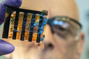
Researchers at the University of Colorado Boulder have unveiled a groundbreaking bioimaging device that operates with significantly lower power and without mechanical parts. This innovation, detailed in a recent study published in Optics Express, holds potential for enhancing the detection of eye and heart conditions.
Revolutionary Non-Mechanical Design
The team of engineers developed a device that utilizes a process called electrowetting to alter the surface shape of a liquid, enabling optical functions. “We are really excited about using one of our devices, in particular for retinal imaging,” said Samuel Gilinsky, the study’s lead author and a recent PhD graduate in electrical engineering. “This could be a critical technique for in-vivo imaging for inside our bodies.”
By eliminating the need for scanning mirrors, the new device requires less electrical power than traditional OCT and bioimaging devices. “The benefits of non-mechanical scanning is that you eliminate the need to physically move objects in your device, which reduces any sources of mechanical failure and increases the overall longevity of the device itself,” Gilinsky explained.
The research team, which includes experts like Juliet Gopinath, Shu-Wei Huang, Victor Bright, Jan Bartos, Eduardo Miscles, and Jonathan Musgrave, emphasizes the importance of creating OCT systems that are compact, lightweight, and safe for human use. “Our work presents an opportunity where we can hopefully detect health conditions earlier and improve the lives of people,” noted Gopinath.
Testing with Zebrafish: A Surprising Ally
To evaluate the device’s biomedical imaging capabilities, the researchers turned to an unexpected subject: zebrafish. These aquatic creatures are often used in OCT research due to the similarity between their eye structure and that of humans. The study focused on identifying the cornea, iris, and retina within the zebrafish.
Achieving in-vivo or other bioimaging requires scientists to identify the structure of samples, such as eyes or internal organs. The research team aimed for benchmarks of 10 microns in axial resolution and around 5 microns in lateral resolution, both smaller than a human hair’s width. “The interesting result was that we were able to actually delineate the cornea and iris in our images,” Gilinsky remarked. “We were able to meet the resolution targets we aimed for, which was exciting.”
This successful testing opens new possibilities for mapping retinal aspects crucial for diagnosing eye conditions like age-related macular degeneration and glaucoma. Furthermore, Gilinsky highlighted the potential for the bioimaging technique to delineate human coronary features, aiding in the diagnosis of heart disease, the leading cause of death in the United States.
Future Prospects and Implications
With expertise in microscopy systems, the research team is optimistic about developing endoscopes that could transform bioimaging technology. “There is a growing push to make endoscopes as small in diameter and flexible as possible to cause as little discomfort as possible,” Gilinsky said. “By using our components, we can maintain a very small-scale optical system compared to a mechanical scanner that can help OCT technologies.”
This project, funded by the Office of Naval Research, National Institutes of Health, and the National Science Foundation, represents a significant step forward in medical imaging technology. As the research progresses, the potential for earlier detection and improved treatment of eye and heart conditions could have profound implications for healthcare worldwide.
The announcement comes at a time when advancements in medical technology are increasingly crucial for addressing global health challenges. As the University of Colorado Boulder’s team continues to refine their device, the promise of more effective diagnostics and enhanced patient outcomes remains on the horizon.