


A groundbreaking artificial intelligence tool developed by researchers at the California NanoSystems Institute at UCLA is set to transform digital pathology. This AI-based system, known as the autonomous quality and hallucination assessment tool for virtually stained tissue images, or AQuA, has demonstrated a remarkable 99.8% accuracy in detecting errors in virtually stained images, a process crucial for diagnosing diseases.
The announcement comes as the medical field increasingly turns to digital solutions to enhance diagnostic accuracy and efficiency. Virtual staining, a technique where AI systems simulate the chemical staining of tissue samples, is at the forefront of this shift. However, these systems can sometimes produce “realistic hallucinations,” errors that can mislead pathologists. AQuA autonomously identifies these errors, even those missed by experienced pathologists, without relying on human-stained slides.
Revolutionizing Traditional Pathology
For over a century, pathologists have relied on staining thin tissue sections with dyes to diagnose diseases such as cancer. This traditional method, while effective, is time-consuming and resource-intensive. According to UCLA researchers, labs can take hours or even a full day to stain samples, whereas AI algorithms can perform virtual staining in under a minute. This not only speeds up the process but also ensures consistency across different regions and laboratories.
Moreover, the environmental impact of traditional staining is significant. The process generates millions of gallons of toxic wastewater annually and consumes costly chemicals. Virtual staining, being carbon neutral, offers a sustainable alternative that could also reduce the need for repeat biopsies by preserving tissue samples.
The Challenge of AI Hallucinations
Despite its advantages, generative AI in pathology is not without risks. Similar to how chatbots may present false information as facts, virtual staining AI can create hallucinations—structures that appear in the stained image but do not exist in the actual tissue sample. These hallucinations can be so convincing that they deceive pathologists, potentially leading to misdiagnoses.
The stakes are particularly high in pathology, where errors can result in unnecessary treatments or missed diagnoses of serious conditions like cancer or organ rejection. As pathology results are critical in formulating treatment plans, ensuring their accuracy is paramount.
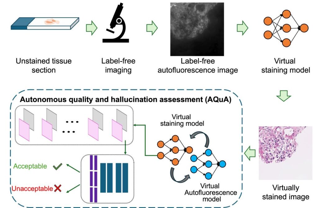
The AQuA Solution
AQuA is designed to mimic the neural networks of the human brain. It evaluates unstained microscopic images, cycling them through a virtual staining algorithm and a reverse process to generate unstained images. Through this method, AQuA learns to distinguish between accurate and hallucinated images, thereby safeguarding against diagnostic errors.
According to sources, AQuA’s ability to detect hallucinations extends beyond its training data, identifying errors even in images stained by human technicians. This capability positions AQuA as a potential gatekeeper in clinical settings, ensuring that pathologists make informed decisions based on reliable images.
Looking Ahead
As virtual staining AI models become more prevalent in clinics, AQuA could play a crucial role in maintaining the integrity of digital pathology workflows. Its applications may include periodic testing and certification of virtual staining models and even protecting against cyberattacks aimed at disrupting healthcare systems.
The study’s corresponding author, Aydogan Ozcan, emphasizes the importance of such innovations. As the Volgenau Professor of Engineering Innovation at UCLA, Ozcan, along with his team, is at the forefront of integrating AI into medical diagnostics. The study, published in Nature Biomedical Engineering, underscores the transformative potential of AI in healthcare.
“The technology described in this study is covered by a patent application filed by the UCLA Technology Development Group on behalf of the Regents of the University of California,” the study notes, highlighting the pioneering nature of this research.
With support from UCLA Samueli’s V. M. Watanabe Excellence in Research Award, the development of AQuA marks a significant step forward in digital pathology, promising a future where diagnostic errors are minimized, and patient outcomes are improved.