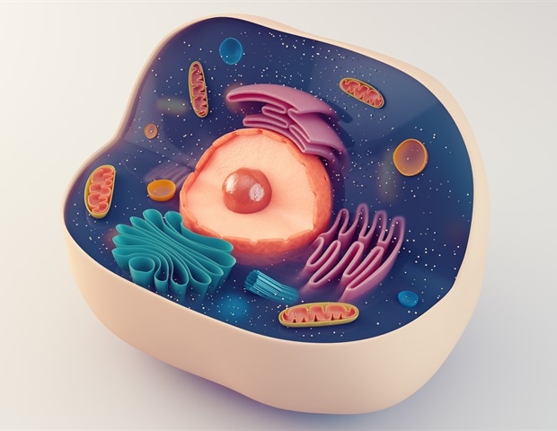
Scientists from leading institutions, including Indiana University and Johns Hopkins Medicine, have unveiled a groundbreaking computer program that mimics human and animal cell behavior. This innovative tool is set to revolutionize the testing and prediction of biological processes, drug responses, and other cellular dynamics, potentially reducing the need for costly live cell experiments.
The program, which could eventually serve as a “digital twin,” is designed to simulate the effects of drugs on cancer and other conditions, as well as gene-environment interactions during brain development. The research, primarily funded by the Jayne Koskinas Ted Giovanis Foundation and the National Institutes of Health, was published in the journal Cell on July 25.
Advancements in Cellular Simulations
According to Dr. Genevieve Stein-O’Brien from Johns Hopkins University School of Medicine, the project began with a workshop on an earlier software version called PhysiCell, developed by Dr. Paul Macklin of Indiana University. PhysiCell uses “agents,” or math robots, to simulate cell behavior based on DNA and RNA rules, allowing researchers to digitally manipulate cell interactions with environmental factors like therapeutics and oxygen.
This approach enables scientists to visualize tumor emergence, therapeutic interactions, and immune system responses, as well as brain cell organization. Dr. Stein-O’Brien’s lab, in collaboration with Dr. Daniel Bergman from the University of Maryland School of Medicine, is further developing the software to simulate brain circuits.
Accessibility and Innovation
Dr. Macklin highlights that while traditional computer modeling programs require extensive math and coding knowledge, the new PhysiCell software introduces a “grammar” that makes it accessible to biologists without programming expertise. “We can now teach scientists to create a basic immunology model in an hour or two,” he notes, emphasizing the program’s potential to model spatial transcriptomics and visualize cell functions in 3D tissue replicas.
Dr. Stein-O’Brien describes the new coding grammar as an “Excel spreadsheet” where each line matches a cell type with a human-readable rule. This setup allows the program to translate these rules into mathematical equations that guide cell behavior, aligning the model with established transcriptome data.
Real-World Applications and Future Potential
Study author David Zhou, a former Johns Hopkins University undergraduate, and Zachary Nicholas, a Ph.D. candidate, contributed to modeling brain development using data from the Allen Brain Atlas. This advancement connects snapshots of cell behavior to create dynamic simulations of cell and tissue interactions over time.
Dr. Elana Fertig, co-leader of the project, explains that cancer cell behavior models were based on data from human pancreatic tumors and mouse experiments. A validation experiment led by Dr. Jeanette Johnson simulated macrophage invasion in breast tumors, revealing that increased EGFR pathway expression promoted cancer growth. This finding was corroborated by live cell observations in the lab.
“We still have a lot of work to do to add more cell behavior data to the program,” says Dr. Johnson, emphasizing the project’s goal of prioritizing hypotheses and therapeutic targets through virtual simulations.
Implications for Medical Research
The development of this software marks a significant step towards a “virtual cell laboratory,” where researchers can prioritize and refine experiments before conducting live tests. Dr. Stein-O’Brien envisions a future where these tools serve as a “digital twin,” enhancing the efficiency and effectiveness of medical research.
Ongoing efforts are focused on integrating artificial intelligence to automate simulation model creation, potentially transforming how digital twin models are developed and applied in medical research.
Collaborative Efforts and Funding
This project was made possible through the collaboration of numerous researchers, including Dr. Stein-O’Brien, Dr. Macklin, and Dr. Fertig, along with contributions from various institutions and funding bodies. The research received financial support from organizations such as the Lustgarten Foundation, the National Foundation for Cancer Research, and the Susan G. Komen Foundation, among others.
As the team continues to enhance the program, the potential for this technology to impact medical research and treatment development remains vast, promising a future where digital simulations play a crucial role in understanding and combating diseases.


