


Caltech scientists have pioneered a groundbreaking method to detect tiny, imperceptible movements on the surface of objects, revealing critical details about what lies beneath. By analyzing the physics of waves traveling across surfaces, whether of manufactured products or the human body, this new technique can determine both the stiffness and thickness of underlying materials or tissues. This innovation lays the foundation for the project’s ultimate goal: enabling inexpensive, at-home health monitoring using little more than a smartphone camera.
“There is information scattered all around us in plain sight that we just haven’t learned to tap into. Our work is trying to leverage that information to recover material properties from inside objects by studying tiny movements on the surface,” says Katie L. Bouman, a professor of computing and mathematical sciences, electrical engineering, and astronomy at Caltech, and both a Rosenberg Scholar and a Heritage Medical Research Institute (HMRI) Investigator.
Revolutionizing Health Monitoring
Bouman and her colleagues from Caltech introduced the technique, known as visual surface wave elastography, and its medical applications in a paper presented at the International Conference on Computer Vision in Honolulu last fall. The lead authors are Alexander C. Ogren (PhD ’24) and Berthy T. Feng (PhD ’25), who completed the work while at Caltech.
Their previous research demonstrated that vibrations captured by a camera could infer varying material properties within a 3D object of known geometry. The motivation was to show that the process could be useful for nondestructive testing, such as detecting internal cracks or verifying the structural integrity of manufactured components. The team has since pivoted towards biomedical applications, where similar surface waves can provide insights into subsurface structures without relying on a pre-existing model of an object’s geometry.
Applications in Medicine
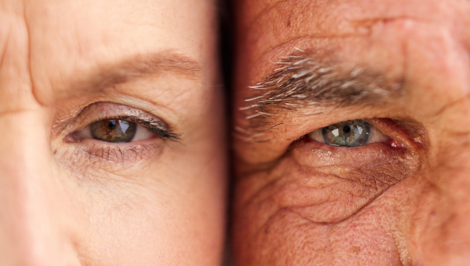
“We asked, ‘Can you infer human tissue properties by the way that motion happens on the skin?’ And the answer is yes,” says Feng, now a postdoctoral fellow at MIT. Visual surface wave elastography enables scientists to measure the stiffness of underlying tissue and its thickness—how far the soft tissue extends before reaching bone. A change in tissue stiffness could serve as a biomarker for diseases such as tumor growth or liver disease. Meanwhile, measurements of thickness could be valuable for monitoring muscle degeneration in diseases that cause atrophy.
“Because we all have cameras in our pockets, we can take frequent, inexpensive measurements of our tissue properties to track our health proactively over time,” Ogren explains. “We could flag concerning changes and nudge you to get it checked out. For example, a system might say, ‘This region of tissue has gotten noticeably stiffer over the last month. You might want to see a doctor for a proper evaluation.'”
Technical Insights and Methodology
The new technique employs an algorithm called phase-based motion processing to detect minute changes in position across the skin, thanks to small-amplitude waves produced by external forces such as quick pressure from a massage gun or sound vibrations from a nearby speaker. The waves could also be excited by vibrations from a wearable device like a watch. Importantly, the method quantifies these movements, which are undetectable by the human eye.
“Basically, the method analyzes local regions of each frame and applies standard signal processing techniques to estimate subpixel motion, resolving shifts as small as one five-hundredth of a pixel,” Bouman explains. The scientists then use spectral analysis to mathematically capture the propagation of those surface waves. The waves are broken down into modes that repeat periodically in time and space, with frequency describing how quickly the mode repeats in time, and wave number describing the spacing between wave peaks and troughs.
“It is exciting to see how powerful computer vision can be in uncovering hidden properties below the surface,” says Chiara Daraio, the G. Bradford Jones Professor of Mechanical Engineering and Applied Physics at Caltech and an HMRI Investigator.
Validation and Future Prospects
In their paper, the team validates its method with data from an anatomically correct simulated human leg and real measurements from a gelatin model. In the gelatin experiments, the new technique yielded results comparable to those of a high-precision instrument called a rheometer. In studies of the simulated leg, the method provided excellent estimates of thickness and stiffness at three different points in the leg, despite the leg having varied nonideal geometry, much like real human bodies.
The research, titled “Visual Surface Wave Elastography,” was supported by funding from the Heritage Medical Research Institute, the Department of Energy, the National Science Foundation, and the Amazon AI4Science Partnership Discovery Grant. In addition to Bouman, Daraio, Ogren, and Feng, Jihoon Ahn (MS ’25) is also an author.
The implications of this research are vast, with the potential to transform how we monitor health conditions at home. As the technology continues to evolve, it could lead to more accessible, affordable, and proactive health care solutions, empowering individuals to take charge of their health with unprecedented ease.