


In a groundbreaking development for breast cancer screening, researchers at MIT have unveiled a miniaturized ultrasound system designed to facilitate more frequent and accessible breast ultrasounds. This innovation is particularly beneficial for individuals at high risk of developing breast cancer, as it enables early detection of tumors, potentially improving treatment outcomes.
The newly developed system comprises a compact ultrasound probe connected to an acquisition and processing module, slightly larger than a smartphone. When linked to a laptop, this portable setup can reconstruct and display wide-angle 3D images in real-time, offering a practical solution for both home and clinical use.
“Everything is more compact, and that can make it easier to be used in rural areas or for people who may have barriers to this kind of technology,” stated Canan Dagdeviren, an associate professor of media arts and sciences at MIT and the senior author of the study. The system’s potential to detect more tumors at an earlier stage could significantly increase the chances of successful treatment.
Frequent Monitoring: A New Era in Breast Cancer Screening
While routine mammograms are the standard for detecting breast tumors, they primarily utilize X-rays and are typically performed annually. However, tumors can develop between these yearly scans, known as interval cancers, which account for 20 to 30 percent of all breast cancer cases and tend to be more aggressive.
Early detection is crucial, as the survival rate for breast cancer diagnosed at its earliest stages is nearly 100 percent. In contrast, this rate plummets to around 25 percent for tumors identified at later stages. The MIT team’s portable ultrasound system aims to supplement regular mammograms, allowing for more frequent monitoring and early detection.
Currently, ultrasounds are usually conducted as follow-ups to mammograms, requiring large, expensive machines and skilled technicians. “You need skilled ultrasound technicians to use those machines, which is a major obstacle to getting ultrasound access to rural communities, or to developing countries where there aren’t as many skilled radiologists,” explained Shrihari Viswanath, an MIT graduate student and co-author of the study.
Technological Advancements: Portable and Cost-Effective Solutions
In 2023, Dagdeviren and her team introduced an array of ultrasound transducers incorporated into a flexible patch, which could be attached to a bra. This allowed users to move an ultrasound tracker along the patch, imaging breast tissue from different angles. However, this setup required connection to a traditional, costly processing machine.
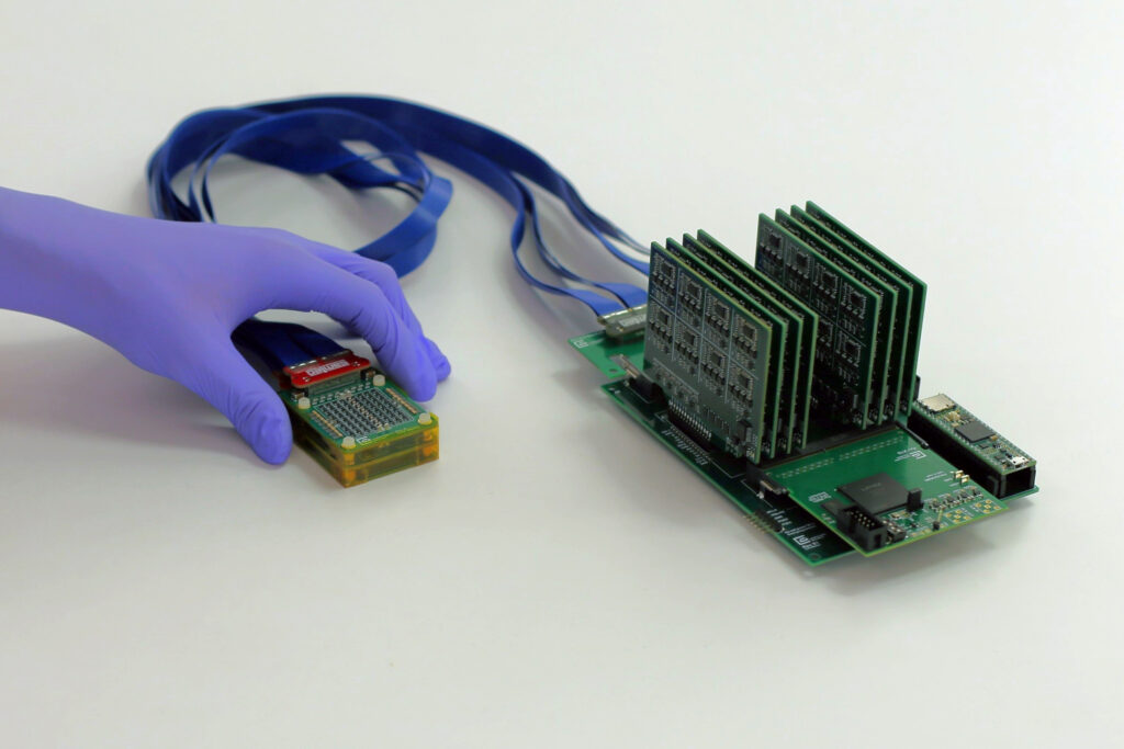
The latest study sought to create a fully portable ultrasound array capable of generating a 3D image of the entire breast by scanning just two or three locations. The new system features a chirped data acquisition system (cDAQ) with an ultrasound probe and a motherboard for data processing. The probe, slightly smaller than a deck of cards, contains an ultrasound array arranged in an empty square shape, enabling 3D imaging of underlying tissue.
The motherboard, slightly larger than a smartphone, costs approximately $300 to produce and uses commercially available electronics. It can be connected to a laptop for image viewing, making the entire system portable. “Traditional 3D ultrasound systems require power expensive and bulky electronics, which limits their use to high-end hospitals and clinics,” noted Anantha Chandrakasan, MIT Provost and co-author of the study.
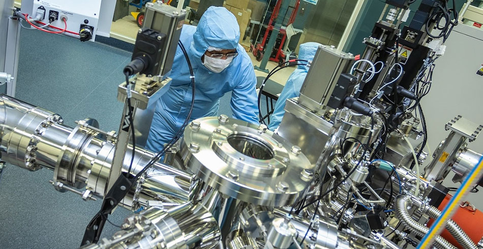
The system’s low power consumption allows it to be powered by a 5V DC supply, such as a battery or small electronic device adapter. “Ultrasound imaging has long been confined to hospitals,” said Md Osman Goni Nayeem, a former MIT postdoc and co-author. “To move ultrasound beyond the hospital setting, we reengineered the entire architecture, introducing a new ultrasound fabrication process, to make the technology both scalable and practical.”
Clinical Trials and Future Developments
The researchers tested the new system on a 71-year-old woman with a history of breast cysts, successfully imaging the cysts and creating a 3D image of the tissue without gaps. The system can image up to 15 centimeters deep and cover the entire breast from just two or three locations. Unlike traditional probes, this device sits on the skin without pressing into the tissue, preventing image distortion.
“With our technology, you simply place it gently on top of the tissue and it can visualize the cysts in their original location and with their original sizes,” Dagdeviren explained. The team is conducting larger clinical trials at the MIT Center for Clinical and Translational Research and Massachusetts General Hospital.
Additionally, the researchers are developing a smaller data processing system, about the size of a fingernail, to connect to a smartphone for image visualization. They plan to create a smartphone app with an AI algorithm to guide users in placing the ultrasound probe optimally. This future version could be incorporated into a wearable sensor for home use by high-risk individuals.
Dagdeviren is working on commercializing the technology with support from various MIT grants and funds. The research received funding from a National Science Foundation CAREER Award, a 3M Non-Tenured Faculty Award, the Lyda Hill Foundation, and the MIT Media Lab Consortium.