


The rising incidence of cancer worldwide has led to an increasing number of surgeries involving the removal of lymph nodes. While these procedures are crucial for cancer staging and preventing the spread of malignancies, they often come with severe long-term consequences. The absence of lymph nodes, which do not naturally regenerate once removed, can lead to secondary lymphedema. This condition manifests as chronic swelling, discomfort, and reduced mobility in affected limbs or regions, severely impacting a patient’s quality of life.
Consequently, a major focus within regenerative medicine is developing strategies to restore or regenerate damaged lymphatic structures to effectively treat secondary lymphedema. Existing approaches have largely centered on stem cells and lymphatic tissue transplantation. However, these techniques often require complex preparation protocols and, more importantly, have demonstrated limited efficacy in improving the key clinical symptoms of lymphedema.
Innovative Technique for Lymphatic Tissue Engineering
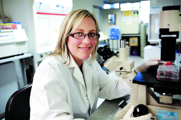
Against this backdrop, a research team led by Associate Professor Kosuke Kusamori from the Faculty of Pharmaceutical Sciences at Tokyo University of Science (TUS), Japan, is pioneering an innovative technique for lymphatic tissue engineering that could revolutionize the treatment of secondary lymphedema. Their study, published in Volume 16 of the journal Nature Communications on November 19, 2025, describes a straightforward protocol to produce bioengineered lymphatic tissues that can restore lymphatic flow after the removal of lymph nodes. This work was co-authored by second-year doctoral student Mr. Shu Obana, Assistant Professor Shoko Itakura, and Professor Makiya Nishikawa, also from TUS.
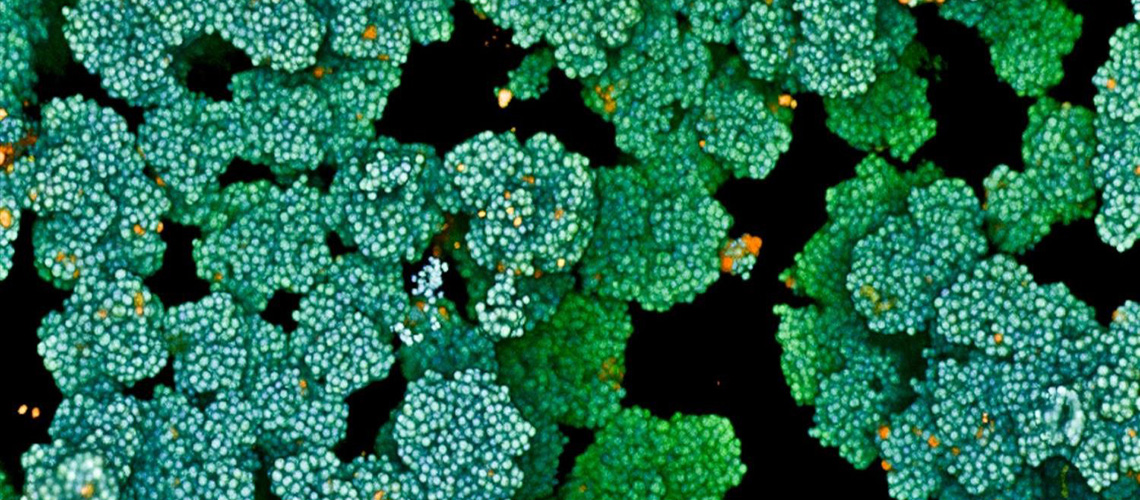
The proposed approach is based on a novel centrifugal cell stacking technique to bioengineer replacement tissue for surgically removed lymph nodes. Initially, mesenchymal stem cells (MSCs), known to support tissue regeneration and provide structural scaffolding, were placed in the wells of a Transwell culture plate. By centrifuging the entire plate, MSCs settled uniformly at the bottom of the wells, forming a first layer. Then, the researchers added lymphatic endothelial cells to the wells, followed by another round of centrifugation to spread them evenly as a second layer. Finally, following a final centrifugation step after adding MSCs again, the result was a three-layered cellular structure, which the researchers called centrifuge-based bioengineered lymphatic tissue (CeLyT).
Promising Results in Animal Models
Using a lymphedema animal model, the team regenerated functional lymph nodes that exhibited structural similarity with native lymph nodes. They confirmed that transplanting CeLyTs restored lymphatic flow in mice whose popliteal and inguinal lymph nodes in the right lower limb had been removed. As a result, these mice exhibited remarkable improvement in lymphedema symptoms, with the thickness of their paws and legs returning to normal within a few weeks. Additionally, mice that received CeLyTs also showed recovery of filtration capacity and immune cell populations such as T cells and macrophages, and lower accumulation of adipose tissue in affected areas, reaching levels similar to normal mice.
The researchers carefully analyzed the structures that formed after CeLyT transplantation to shed light on the observed therapeutic effects.
“CeLyTs may initially induce lymph and blood vessel formation around the transplantation site, leading to the establishment of an immature lymph node-like structure formed by incorporating host-derived cells into the tissue within several days, followed by its maturation and ability to function as a lymph node within 10 days after transplantation,” explains Dr. Kusamori.
The Future of Lymphedema Treatment
This study marks the world’s first successful regeneration of fully functional lymph nodes through cell transplantation, offering a promising therapeutic option for patients who develop lymphedema following oncologic surgeries involving lymph node dissection. Economically, a single transplantation could provide long-lasting therapeutic benefits, substantially reducing the cumulative costs associated with repeated hospital visits and long-term use of compression garments. Overall, these results highlight the strong curative potential of introducing appropriately bioengineered tissue into the lymphatic system, surpassing the efficacy of current treatment options for lymphedema.
“Although compression therapy represents the gold standard for the treatment of lymphedema in clinical practice, it usually delays the swelling in the paws of lymphedema mice. By contrast, CeLyTs were more effective at suppressing lymphedema, also exhibiting strong therapeutic effects even in a more severe chronic lymphedema model,” remarks Dr. Kusamori. “Moreover, CeLyTs demonstrated a greater lymphedema-suppressive effect, compared with bioengineered tissues fabricated by other tissue engineering methods.”
The move represents a significant leap forward in the treatment of lymphedema, offering hope for future clinical applications. As researchers continue to refine and test this technique, the potential for improving the quality of life for countless patients becomes increasingly tangible. With further studies and eventual clinical trials, CeLyTs could soon become a standard treatment, transforming the landscape of lymphedema care.