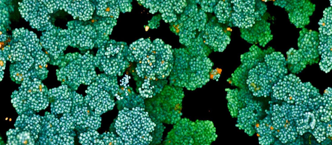


In the realm of radiation therapy, precision is not just a goal; it is a lifesaving necessity. Oncologists meticulously map the size and location of a tumor to deliver high-dose radiation that eradicates cancerous cells while preserving healthy tissue. This meticulous process, known as tumor segmentation, traditionally relies on manual efforts, which can be time-consuming, vary between practitioners, and occasionally result in critical tumor areas being overlooked.
Now, a groundbreaking development by a team of Northwestern Medicine scientists promises to transform this process. They have introduced an artificial intelligence tool named iSeg, which not only matches doctors in accurately outlining lung tumors on CT scans but also identifies regions that may be missed by some physicians. This advancement is highlighted in a comprehensive new study.
Unlike previous AI technologies that were limited to static images, iSeg is the pioneering 3D deep learning tool capable of segmenting tumors as they move with each breath—a vital aspect in planning radiation treatments, which are administered to half of all cancer patients in the United States during their treatment journey.
“We’re one step closer to cancer treatments that are even more precise than any of us imagined just a decade ago,” said Dr. Mohamed Abazeed, senior author and chair of radiation oncology at Northwestern University Feinberg School of Medicine.
Building and Testing iSeg
The creation of iSeg involved training the AI using CT scans and physician-drawn tumor outlines from hundreds of lung cancer patients across nine clinics within the Northwestern Medicine and Cleveland Clinic health systems. This extensive dataset surpasses the smaller, single-hospital datasets that have been the norm in many past studies.
Upon completion of its training, iSeg was tested on patient scans it had never encountered before. The AI’s tumor outlines were then compared to those drawn by experienced physicians. The study revealed that iSeg consistently matched expert outlines across various hospitals and scan types. Moreover, it flagged additional areas that some doctors missed, which were associated with poorer outcomes if left untreated. This suggests that iSeg may help identify high-risk regions that often go unnoticed.
“Accurate tumor targeting is the foundation of safe and effective radiation therapy, where even small errors in targeting can impact tumor control or cause unnecessary toxicity,” Abazeed emphasized.
Sagnik Sarkar, the study’s first author and a senior research technologist at Feinberg, added, “By automating and standardizing tumor contouring, our AI tool can help reduce delays, ensure fairness across hospitals, and potentially identify areas that doctors might miss—ultimately improving patient care and clinical outcomes.”
Potential for Clinical Deployment
The research team is currently testing iSeg in clinical environments, directly comparing its performance to that of physicians in real time. They are also integrating features such as user feedback and expanding the technology to other tumor types, including liver, brain, and prostate cancers. Additionally, plans are underway to adapt iSeg for use with other imaging methods, such as MRI and PET scans.
“We envision this as a foundational tool that could standardize and enhance how tumors are targeted in radiation oncology, especially in settings where access to subspecialty expertise is limited,” stated co-author Troy Teo, an instructor of radiation oncology at Feinberg.
Teo further noted, “This technology can help support more consistent care across institutions, and we believe clinical deployment could be possible within a couple of years.”
Implications and Future Directions
The introduction of iSeg represents a significant leap forward in the field of radiation oncology. By providing a tool that can accurately and consistently delineate tumors, the AI has the potential to improve treatment outcomes and reduce the variability inherent in manual tumor segmentation. This advancement is particularly crucial in areas with limited access to specialized oncology expertise.
As the research team continues to refine and test iSeg, the potential for widespread clinical adoption grows. The ability to adapt the tool to various imaging modalities and tumor types further broadens its applicability, promising to enhance the precision and effectiveness of cancer treatments across the board.
With the study published in the journal npj Precision Oncology, the medical community eagerly anticipates the next steps in the development and deployment of this transformative technology. The future of cancer treatment may very well be shaped by the integration of AI tools like iSeg, offering hope for more personalized and effective care for patients worldwide.