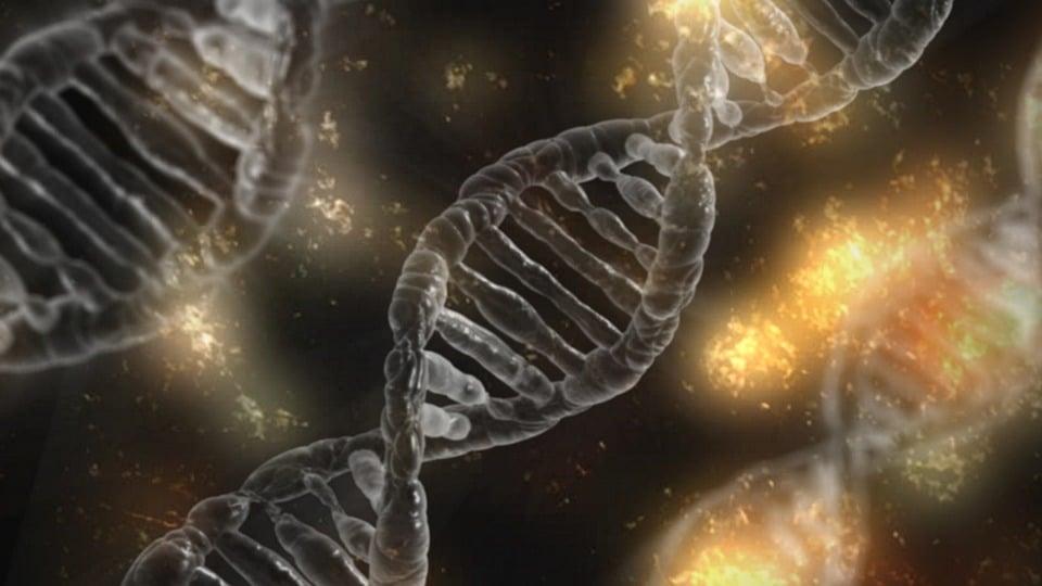


NIMS, in collaboration with Nagoya University, Gifu University, and the University of Adelaide, has unveiled a groundbreaking method for simultaneously imaging DNA and RNA within cells using harmless infrared to near-infrared light. This innovative study, published in Science Advances on October 23, 2025, marks a significant advancement in the field of cellular imaging, enabling high-precision detection of all stages of cell death. This breakthrough paves the way for early detection of cell aging and damage, crucial for disease prevention.
The announcement comes as researchers continue to seek methods that can safely and effectively monitor cellular changes throughout their life cycle. The ability to observe these changes is essential for developing therapeutic strategies for various diseases. However, existing imaging techniques often fall short, particularly in ultraviolet-visible (UV-vis)–sensitive cells, where they fail to capture early responses or distinguish multiple injury states. These limitations can lead to delayed diagnoses and an incomplete understanding of cellular fate post-treatment.
Revolutionary Imaging Technique
The research team has successfully visualized both DNA and RNA inside living cells by employing two types of harmless excitation light alongside fluorescent dye probes known as N-heteroacene dyes. These dyes bind differently to DNA and RNA, allowing for simultaneous and safe imaging. This dual imaging approach not only assesses sustained DNA damage but also reveals that RNA imaging provides higher sensitivity for predicting early stages of cell damage and aging.
“This dual DNA/RNA imaging enables early evaluation of cellular damage and precise detection of all four stages of cell death,” the researchers noted. “The approach surpasses the limitations of current imaging systems by enabling visualization of single-cell state transitions.”
This development follows a growing need for universal, highly sensitive imaging methods that utilize harmless infrared to near-infrared excitation light. Such methods are crucial for safely monitoring complete cell states, and this new technique opens up possibilities for ultra-early detection of cellular damage and aging, non-toxic live-cell diagnostics, and high-throughput drug screening workflows.
Implications for Medical Research
According to experts, this advancement could revolutionize the field of medical research and diagnostics. By providing a means to detect cellular changes at an earlier stage, researchers can better understand the progression of diseases and develop more effective treatment strategies.
Dr. Emily Chen, a leading researcher in cellular imaging, commented, “This method represents a significant leap forward. The ability to observe both DNA and RNA simultaneously in living cells without causing harm is a game-changer for our understanding of cellular processes and disease progression.”
By the Numbers: The study’s method allows for the precise detection of all four stages of cell death, a capability that current imaging systems lack.
Future Prospects and Applications
Looking ahead, the research team plans to apply this method to living organisms in future studies. Their goal is to establish techniques for early disease detection, cellular stress monitoring, and precision medical strategies. Ultimately, they hope to develop technologies that can determine a “pre-disease” state—when a person is drifting away from health—by simply observing their cells.
Meanwhile, the implications of this research extend beyond disease prevention. The ability to conduct non-toxic live-cell diagnostics and high-throughput drug screening could accelerate the development of new pharmaceuticals and therapies, offering hope for patients with currently untreatable conditions.
The move represents a significant step forward in the quest for safer and more effective cellular imaging techniques. As researchers continue to explore the potential of this method, the possibilities for its application in both clinical and research settings appear promising.
As the scientific community awaits further developments, this breakthrough in DNA and RNA imaging stands as a testament to the power of collaboration and innovation in advancing our understanding of cellular biology and its implications for human health.