


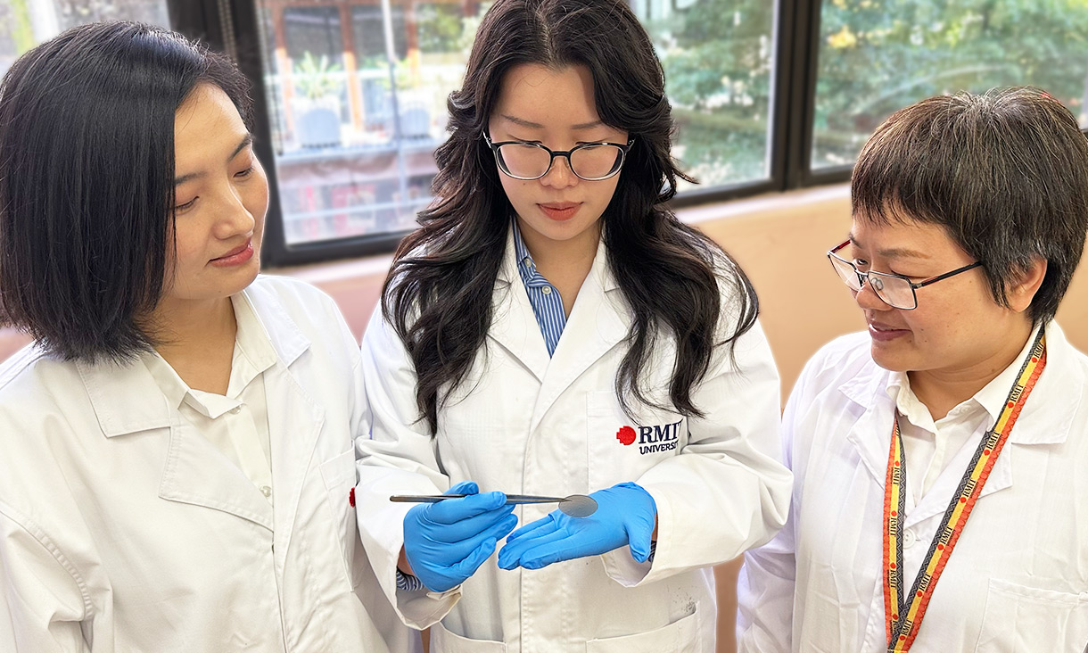
A groundbreaking study published in the Journal of Orthopaedic Research introduces a novel, noninvasive method to assess the health of the Achilles tendon in professional ballet dancers. Utilizing advanced imaging techniques, this method holds promise for preventing injuries and refining rehabilitation strategies not only for athletes but also for the broader public.
The research employs a combination of multi-echo ultrashort echo time (UTE) magnetic resonance imaging (MRI) and shear wave elastography (SWE) ultrasound to evaluate the structural and functional aspects of the Achilles tendon. This dual approach allows for a comprehensive assessment of tendon health, particularly focusing on collagen and other vital components.
Understanding the Technique
Multi-echo UTE MRI provides a detailed view of the tendon’s structure by capturing images that highlight the organization of collagen fibers. Meanwhile, SWE ultrasound offers a functional perspective by measuring tendon stiffness, a critical factor in assessing tendon health. The integration of these techniques offers a holistic view of the tendon, correlating structural integrity with functional capacity.
According to the study, professional dancers exhibited greater tendon stiffness compared to non-dancers. This observation aligns with previous findings that suggest repeated physical loading during training enhances tendon stiffness. The UTE MRI results were consistent with the stiffness measurements obtained through SWE ultrasound, reinforcing the reliability of this combined approach.
Implications for Injury Prevention and Rehabilitation
The implications of this study are significant for both injury prevention and rehabilitation. By providing a clearer picture of tendon health, this method could lead to more targeted strategies for preventing injuries in high-risk groups such as athletes and dancers. Additionally, it could inform rehabilitation protocols, ensuring they are tailored to the specific needs of individuals based on detailed assessments of their tendon health.
“These findings highlight the potential of integrating UTE and SWE imaging to investigate tendon structure‐function relationships and adaptations to mechanical loading,” the authors of the study noted. “Enhanced structure‐function assessment of tendon health and injury status could improve rehabilitation protocols or injury prevention strategies for athletes, including professional dancers.”
Broader Applications and Future Research
While the study focuses on professional ballet dancers, the applications of this technique extend far beyond the dance studio. The method could be adapted for use in various sports disciplines, potentially revolutionizing how tendon health is monitored and managed across different athletic populations. Moreover, the general public could benefit from these advancements, particularly those engaged in regular physical activity.
Looking ahead, further research is needed to refine these imaging techniques and explore their full potential. Future studies could investigate how these methods can be integrated into routine health assessments and explore their effectiveness in predicting and preventing tendon-related injuries.
The study, accessible via the Journal of Orthopaedic Research, marks a significant step forward in sports medicine and rehabilitation. As researchers continue to explore the capabilities of UTE MRI and SWE ultrasound, the potential for improving athlete care and injury prevention remains vast.
In conclusion, this innovative approach to assessing Achilles tendon health represents a promising advancement in both the prevention and treatment of tendon injuries. As the technique evolves, it may become a standard tool in the arsenal of sports medicine professionals, enhancing the well-being of athletes and active individuals alike.
