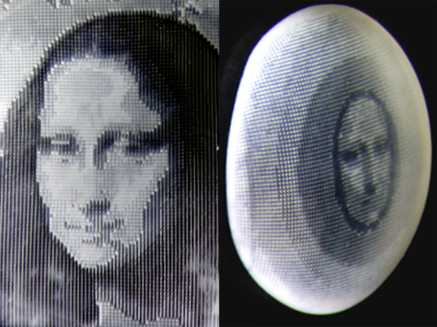


The Beaker Street Science Photography Prize has once again captivated audiences with its stunning visual exploration of the microscopic world. This year’s standout entry, titled “Inner Terrain,” utilizes advanced microscopy to reveal the hidden complexities of human health. Captured by photographer Kelli Miller, the image provides a fascinating glimpse into the intricate structures found during a dried blood evaluation.
At the heart of this captivating image are polymerised protein puddles (PPPs), which appear as white areas resembling holes, with black tentacle-like lines extending from their forms. These soft clots vary in size, ranging from small white dots to larger formations, and are indicative of the tissue’s health. Often associated with free radical damage, oxidative stress, and toxins, PPPs offer a detailed insight into the individual’s overall health condition.
The Science Behind the Image
The use of microscopy in health evaluations is a growing field, providing unparalleled insights into the body’s internal state. The dried blood evaluation technique, employed in Miller’s photograph, is a method that allows for the detection of various health markers. By examining the blood’s microscopic structure, scientists can assess the presence of oxidative stress and other potential health issues.
According to Dr. Emily Carter, a leading expert in microscopy and blood analysis, “The presence of PPPs can be a crucial indicator of underlying health problems. These formations can reveal the extent of oxidative damage and help in diagnosing potential issues before they manifest more severely.”
Historical Context and Advances
Microscopy has long been a cornerstone of scientific discovery, dating back to the invention of the first compound microscope in the late 16th century. Over the centuries, advancements in technology have allowed researchers to delve deeper into the microscopic world, unveiling details that were once invisible to the naked eye.
The Beaker Street Science Photography Prize celebrates these advancements by showcasing images that not only highlight the beauty of science but also its potential to transform our understanding of health and disease. “Inner Terrain” is a testament to how far microscopy has come, offering both aesthetic appeal and scientific value.
Implications for Health and Research
The implications of findings like those depicted in Miller’s photograph are significant. As researchers continue to explore the microscopic realm, the potential for early disease detection and prevention grows. By identifying markers such as PPPs, healthcare providers can develop more targeted treatment plans, ultimately improving patient outcomes.
Dr. Carter emphasizes the importance of continued research in this field, stating, “The more we understand about the microscopic indicators of health, the better equipped we are to tackle diseases at their root. This kind of research is crucial for the future of preventative medicine.”
Meanwhile, the Beaker Street Science Photography Prize continues to inspire both scientists and the public alike, bridging the gap between art and science. As the competition evolves, it serves as a reminder of the endless possibilities that lie within the microscopic world.
Looking ahead, the integration of advanced imaging techniques in routine health assessments could revolutionize how we approach personal healthcare. As technology progresses, the ability to visualize and interpret the body’s inner workings will undoubtedly lead to more personalized and effective medical interventions.
In conclusion, the Beaker Street Science Photography Prize not only celebrates the beauty of scientific discovery but also underscores the critical role of microscopy in advancing our understanding of health. As we continue to explore the unseen, the potential for transformative breakthroughs remains vast and promising.